Knowledge of the structural organization of the nervous system is essential to the proper understanding of its normal, as well as altered, function.
For didactic purposes, the nervous system is generally divided into central and peripheral components. The central nervous system includes the brain and the spinal cord. The peripheral nervous system includes cranial and peripheral nerves and associated ganglia. The autonomic nervous system includes parts of the central and peripheral nervous systems.
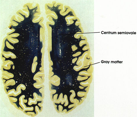
The brain includes the cerebral hemispheres, the cerebellum, and the brain stem. The latter includes the diencephalon; mesencephalon, or midbrain; pons; and medulla oblongata. Each of these components is made up of cell groups and fibers, arranged in a manner that characterizes the particular component. Bundles of nerve fibers serving a common function and sharing a common origin and destination are grouped together in tracts or fasciculi. A group of neurons serving a common function forms a nucleus.
Knowledge of the existence and location of tracts has been gained through years of clinical observation and experimentation in both animals and man. Some of the methods used in the tracing of neural pathways follow:
-
Study of normal preparations: Many aspects of fiber connectivity of the nervous system have been elucidated by early studies using normal material and methods that demonstrate myelin sheaths (Weigert and Weil methods) or that impregnate cell bodies and their processes (Golgi method). The disadvantage of these methods is the difficulty of determining the site of termination of these fibers.
-
Myelinogenesis: This method, introduced by Flechsig, makes use of the fact that different fiber tracts become myelinated at different times in their development. Thus, study of the nervous system in embryos and in early neonatal life often affords information about the existence and locality of the different fiber tracts. This method is infrequently used today.
-
Study of pathological conditions in man and experimental lesions in animals: This method accounts for most of our current knowledge of neural connectivity. Although human material has been of use, experimentally produced lesions in animals have the major advantage of selectivity of site and size. Caution should be exercised, however, in applying to humans results achieved in experimental animals.
After a lesion has been produced in animals or man and sufficient time has elapsed for anterograde degeneration to set in, the brains and spinal cords can be studied, and degenerated tracts can be localized by one of the following three methods:
-
Methods that stain normal myelin (Weigert, Weil): In such preparations, normal myelin appears dark blue or black, and the degenerated tracts will be conspicuous by their failure to pick up the stain.
-
Methods that stain degenerating myelin (Marchi): In such preparations, only degenerating myelinated tracts pick up the stain and can be followed from origin to termination. Normal myelinated tracts remain unstained. A major advantage of the Marchi method is that positive results may be obtained years after degeneration has occurred, making it particularly useful in the study of human material postmortem. One disadvantage of this method is that thinly myelinated or unmyelinated tracts will not stain. Another disadvantage is that it does not stain degenerating terminals; hence, the exact site of termination of a tract cannot be determined with certainty.
-
Methods that stain degenerating axons (Nauta-Gygax, Fink-Heimer, De Olmos): These are silver impregnation techniques that stain degenerating axons and pre-terminals (Nauta-Gygax) or terminals (Fink-Heimer, De Olmos). These methods have a distinct advantage over myelin methods, since they are capable of revealing poorly myelinated as well as unmyelinated nerve fibers, because the axon, and not the myelin, is stained by these methods.
A neuroanatomist is interested not only in the location and course of fiber tracts but also in their site of termination. To determine the latter, methods that stain the terminal boutons (Glees, Bodian) are used. Electron microscopy can also be used for this purpose.
-
Retrograde cell changes: By this method, the position of neurons giving rise to the tract is determined. Such neurons undergo chromatolytic changes of their Nissl substance or disappear completely (retrograde degeneration) if their axon is severed. These changes can be demonstrated by any of the methods that stain ribonucleic acids (Nissl material), the Nissl stains.
-
Autoradiography: This is a relatively recent pathway tracing technique used in brain research. it utilizes the principle that radioactive amino acids injected in the vicinity of neuronal perikarya will be taken up by the neuron, incorporated into its macromolecules, and transported anterograde along the axon to its terminal. After a finite time following injection, the radioactive amino acid can be demonstrated by autoradiography. By this method, the path of a neural tract can be traced from its origin to its termination.
-
Enzymatic method: When the enzyme horseradish peroxidase (HRP) is injected at the site of termination of nerve fibers, it is taken up by the nerve terminals and transported retrograde to the perikaryon where it is visualized by an enzyme histochernical technique as brown granules in the soma and dendrites.
-
Fluorescence method: This method, introduced in the early 1960s, is used to trace the fiber pathways of adrenergic and monaminergic neural systems. It relies on the observation that primary amines form fluorescent condensation products when treated with formaldehyde in the presence of protein. Fluorescent condensation products are demonstrated in cells, axons, and terminals by fluorescence microscopy.
-
Physiological exploration: By this method, stimulation and recording techniques are used to establish the presence or absence of structural and/or functional relationships between two or more loci in the nervous system. The stimulation and recording of evoked potentials may be orthodromic (recording of activity in the terminal projection site of a fiber system) or antidromic (recording of activity in the cells of origin when their axon terminals or axons are stimulated). Gross stimulation and recording techniques reflect the relationship between groups of neurons; intracellular recordings reflect the relationship between pairs of neurons.
These methods used to study neural connectivity are based on the principle of the neuron as a trophic unit. If an axon is transected, its peripheral parts, including its termination, undergo degeneration. This is referred to as anterograde degeneration. The methods described in 3 above are used to show this type of degeneration. Simultaneously with anterograde degeneration, changes occur in the proximal components of the neuron, namely in the proximal axon, the cell body, and dendrites. These changes are known as retrograde changes.
When considered together, anterograde and retrograde methods allow a detailed mapping of neural connectivity.
SPINAL CORD
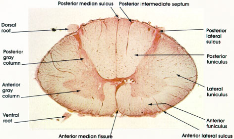
Cervical region
Human, Müller's fluid, carmine stain, 8 x.
Dorsal root: The dorsal root carries both myelinated and unmyelinated afferent fibers to the spinal cord. Each fiber is the central process of a dorsal root ganglion cell.
Posterior gray column: Long and narrow column of gray matter reaches almost to the surface of the spinal cord. Primarily concerned with sensory input.
Anterior gray column: Short and broad column of gray matter. Concerned with motor function. Both posterior and anterior gray columns are sites where sensory and motor cell bodies, respectively, are found.
Ventral root: Bundle of somatic motor fibers (axons of somatic motor neurons) and preganglionic fibers (axons of autonomic motor neurons). Constitute the efferent outflow of the spinal cord.
Anterior median fissure: About 3 mm deep. Contains blood vessels (anterior spinal artery) supplying the anterior two thirds of the cord.
Anterior lateral sulcus: Site of exit of ventral root. Hardly distinguishable in this preparation.
Anterior funiculus: Between the anterior median fissure and anterolateral sulcus (ventral root). Merges with the lateral funiculus. Contains ascending and descending tracts.
Lateral funiculus: Between the dorsal and ventral roots. Merges with the anterior funiculus. Contains ascending and descending tracts.
Posterior lateral sulcus: Site of entry of dorsal root.
Posterior funiculus: Between posterior median sulcus and dorsal root. Contains ascending tracts. Posterior intermediate septum: Found only in cervical and upper thoracic segments. Posterior median sullcus: About 5 mm deep, reaches the deep-lying gray matter.
SPINAL CORD
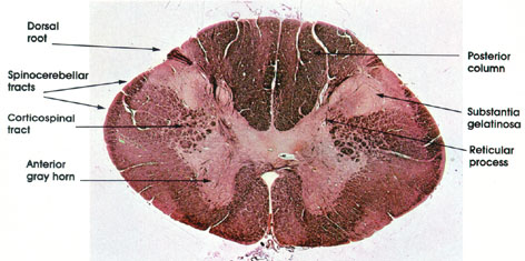
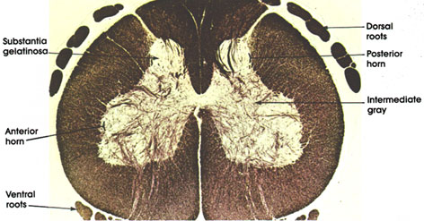
Cervical level
Human, 10% formalin, Weigert-carmine, 11 x.
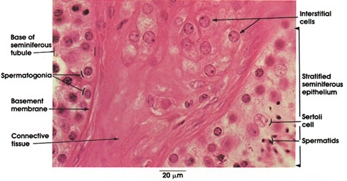
Posterior column: The white matter located between the posterior median septum and the medial border of the posterior horn. Contains heavily myelinated nerve fibers that form the ascending gracile and cuneate tracts. These tracts carry impulses from proprioceptive and tactile receptors (Pacinian corpuscles, Golgi tendon organs, neuromuscular spindles, and Meissner's corpuscles), which give rise to sensations of position, movement, and tactile localization.
Substantia gelatinosa: Appears as a cap-like structure at the head of the posterior horn. It is largest in the first cervical and in the lumbosacral segments but extends the whole length of the cord. This nucleus contains small cells about 6 to 20 µm in diameter and is the primary associative center of the posterior horn for incoming impulses carried by the dorsal root. This nucleus is an important part of the pathway for pain, temperature, and some tactile impulses.
Dorsal root: Sensory nerve fibers entering the spinal cord. Follow some of these fibers to the anterior horn where they terminate on motor neurons.
Spinocerebellar tracts: The spinocerebellar tracts convey impulses to the cerebellum from muscle, tendon, and joint proprioceptors, thus enabling the cerebellum to coordinate skeletal muscle activity (posture and movement).
Reticular process: Contains small and medium-sized cells, which send their axons to the adjacent as well as to the opposite anterolateral white column. Also known as the nucleus reticularis.
Anterior gray horn: Contains cells whose axons pass to the extrafusal fibers of striated skeletal muscle. The cells are the largest found in the spinal cord. These multipolar neurons have as many as 20 dendrites, and axons approximately 12 µm in diameter. Smaller neurons with thin axons (gamma efferent fibers) supply the intrafusal muscle fibers of the neuromuscular spindle
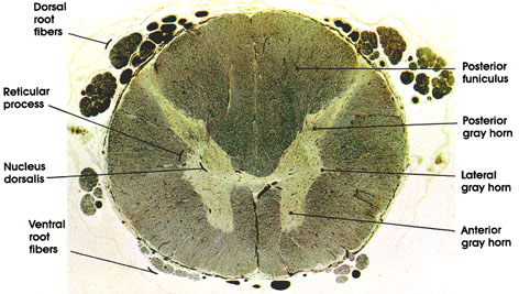
SPINAL CORD
Thoracic region
Human, Müller's fluid, Weigert's method, 10 x.
Dorsal root fibers: Central processes of dorsal root ganglia.
Reticular process: Characteristic of cervical levels of the spinal cord but also located at other spinal levels. Located between the posterior and anterior horns and produced by an extension of gray matter into the adjacent white substance. Constitutes the lateral zone of lamina V of Rexed.
Nucleus dorsallis: Distinct nuclear mass located in the medial part of the base of the posterior horn. In this nucleus, dorsal root fibers synapse with neurons destined to form the dorsal (posterior) spinocerebellar tract. The nucleus extends between C8 and L2 spinal segments. Also known as the column of Clarke*.
Ventral root fibers.
Posterior funiculus: The white matter of the cord located between the posterior central (median) septum and the medial border of the posterior horn. Contains heavily myelinated fibers that form the gracile and cuneate tracts. Note the large size of this funiculus at this level compared to lower levels of the spinal cord. .
Posterior gray horn: A mass of neurons in the posterolateral part of the spinal cord. Receive collaterals or terminals of dorsal root fibers. Sends axons to anterior horn cells, interneurons, or to ascending tracts.
Lateral gray horn: Characteristic of thoracic level, this projection is formed by the intermediolateral nucleus. Contains visceral efferent neurons of the sympathetic nervous system. Extends from C8 to L2-4. Axons of neurons here exit with the ventral horn fibers to terminate in the chain of ganglia (sympathetic), where they synapse with ganglion cells whose axons are widely distributed to the iris (dilator smooth muscle); lacrimal, salivary and sweat glands; bronchi; heart; smooth muscle of the gastrointestinal tract; sex organs; urinary bladder; adrenal medulla; and blood vessels.
Anterior gray horn: A mass of large neurons in the anterolateral part of the spinal cord. Contains somatic efferent neurons. Compare the size of the horn at this level with those seen at higher and lower levels.
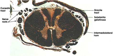
SPINAL CORD LESION
Thoracic level dorsal funiculus
Human, 10% formalin, Weigert-carmine, 9.4 x.
Gracile tract: Note that the gracile tract stains lighter than the adjacent cuneate tract. See Plates 317 and 322. This is due to the degenerating myelinated fibers in the gracile tract, which are not stained by this method. This slide was taken from a human with a spinal cord lesion in the dorsal (posterior) funiculus below the sixth thoracic segment. Fibers entering the spinal cord below this level form the gracile tract. The fibers entering above T6 form the cuneate tract and therefore escape degeneration.
Substantia gelatinosa: Contains 6 to 20 µm diameter neurons. Note small size compared with that of cervical cord. Concerned with sensory relay and integration.
Intermediolateral horn: Lateral gray horn. Characteristic of dorsal (thoracic) and upper lumbar segments. Contains sympathetic neurons.
Nerve roots: Incoming dorsal roots.
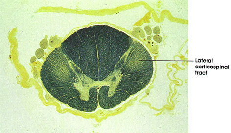
SPINAL CORD
Primary lateral sclerosis
Human, 10% formalin, Weil, 8 x.
This is a section of the spinal cord stained with Weil's method. This method stains normal myelinated fiber tracts black. The lightly stained lateral corticospinal tract stands out in contrast to the darkly stained normal myelin elsewhere in the spinal cord. This selective loss of myelinated fibers in the lateral corticospinal tract is characteristic of a spinal cord disorder known as primary lateral sclerosis. Patients afflicted with this disorder show all the signs of upper motor neuron lesion, including weakness, spasticity, hyperreflexia, Babinski* reflex, and clonus.
SPINAL CORD
Lumbar region
Human, 10% formalin, Well, 8 x.
Dorsal roots: Central processes of dorsal root ganglion cells. Convey afferent (sensory) impulses to the spinal cord from peripheral receptor organs..
Substantia gelatinosa: Cap-like structure at the head of the posterior horn. Extends the whole length of the cord. Contains small neurons about 6 to 20 µm in diameter and is the primary associative center of the posterior horn for incoming impulses carried by the dorsal root. This nuclear mass is an important part of the pathway for pain, temperature, and some tactile impulses.
Dorsal root fiber collaterals: Heavily myelinated dorsal root fiber collaterals that enter the spinal cord to modify pain transmission or establish segmental reflexes.
Anterior funiculus: Between the anterior median fissure and anterolateral sulcus (ventral roots). Merges with the lateral funiculus. Contains several ascending and descending tracts.
Ventral roots: Bundles of somatic motor fibers (axons of somatic motor neurons in the anterior horn). Constitute the efferent outflow of the spinal cord.
Anterior gray horn: A mass of large multipolar motor neurons and interneurons. Axons of motor neurons form the ventral roots. Compare size of anterior gray horn at this level with those seen at higher and lower levels.
Lateral funiculus: Between the dorsal and ventral roots. Merges with the anterior funiculus. Contains major ascending and descending fiber tracts, including the lateral corticospinal, spinothalamic, and spinocerebellar tracts.
Posterior funiculus (fasciculus gracilis): The posterior funiculus at this level contains the fasciculus gracilis only, whereas at higher levels (above the sixth thoracic spinal segment), it contains the fasciculi gracilis and cuneatus. Compare size of the posterior funiculus at this level with that at levels below and above
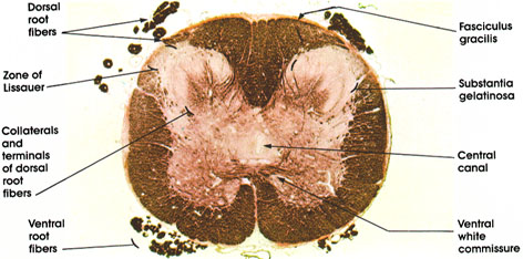
SPINAL CORD
Sacral region
Human, Müller's fluid, Weigert's method with carmalum stain, 14 x.
Dorsal root fibers: Bundles of heavily myelinated nerve fibers entering the spinal cord. Represent central processes of dorsal root ganglion neurons. Convey afferent impulses from peripheral organs to the spinal cord. Some of these fibers go directly to form tracts (fasciculus gracilis), others give collaterals or terminate on neurons in the spinal cord.
Zone of Lissauer*: Also known as fasciculus dorsolateralis. Composed of fine myelinated and non- myelinated fibers that carry pain, thermal, and light touch impulses or that interconnect different levels of the substantia gelatinosa.
Ventral root fibers: Axons of somatic, fusimotor, and visceral motor neurons in the anterior (ventral) and lateral gray columns. Heavily myelinated.
Fasciculus gracilis: Heavily myelinated ascending fiber system. Conveys kinesthetic sense and discriminative touch. Note the absence of the fasciculus cuneatus, which appears at spinal cord levels above T6.
Substantia gelatinosa: An expanded cell mass that forms the cap of the posterior gray horn of the spinal cord. Its size is related to that of the dorsal root. This area functions as an association region for incoming impulses. This region corresponds to lamina II of Rexed.
Central canal: Runs throughout the length of the cord. Partially obliterated in the adult.
Ventral white commissure: Bundle of myelinated fibers crossing from one side of the spinal cord to the other.
SPINAL CORD
Dorsal root collaterals coronal
Dog, Müller's fluid, Weigert's method, 3.75 x.
This is a section of the spinal cord showing the distribution of dorsal root collaterals. Note that the coarser, heavily myelinated collaterals are medially located. They are seen passing to the posterior horn, intermediate gray, and anterior horn. Many fibers of this bundle enter the posterior funiculus. Finer, poorly myelinated collaterals are more laterally located and are seen entering the substantia gelatinosa. Some heavily myelinated fibers also enter the substantia gelatinosa. The medial bundle is the larger of the two. Collaterals that pass directly to the anterior horn constitute components of monosynaptic reflex arc. Their number is relatively small since most of the collaterals to the anterior horn terminate on at least one interneuron before reaching the final efferent neuron. Note the bundle of motor fibers (axons of motor neurons) leaving the anterior horn to form the ventral roots.
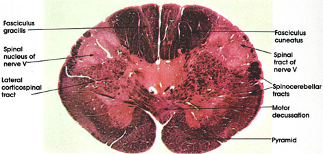
MEDULLA OBLONGATA
Motor decussation coronal
Human, 10% formalin, Pal-Weigert and carmine stains, 11 x.
Fasciculus gracilis: Rostral continuation of the same tract seen at several spinal cord levels The lightly stained islands within the fasciculus represent neurons of the nucleus gracilis. This nucleus is larger at more rostral levels.
Fasciculus cuneatus: Rostral continuation of the same tract seen at spinal cord levels .
Spinal nucleus of nerve V: Functionally analogous to and structurally a continuation of the substantia gelatinosa seen at several spinal cord levels. In it terminate fibers of the descending (spinal) tract of cranial nerve V (trigeminal), which enters the neuraxis at a rostral level. The nucleus is primarily concerned with the perception of pain and thermal sense from the homolateral face.
Spinal tract of nerve V: Thinly myelinated fibers, hence less densely stained than the heavily myelinated fibers of the fasciculi gracilis and cuneatus or the spinocerebellar tracts. This tract is composed of descending trigeminal fibers and extends from the site of entry of the trigeminal nerve in the pons down to at least the second cervical spinal segment. Primarily concerned with pain and thermal sense. Synapse in the spinal nucleus of nerve V.
Spinocerebellar tracts: Heavily myelinated. Continuation of the same tracts seen at several spinal cord levels on their way to the cerebellum.
Motor decussation: Constitutes one of the most conspicuous features of sections at this level. Site of crossing of the pyramids to form the lateral corticospinal tracts. Approximately 75 to 90 per cent of descending pyramidal fibers cross at this level. The motor decussation forms the basis for voluntary motor control of one half of the body by the contralateral cerebral hemisphere.
Lateral corticospinal tract: Formed by decussation of the pyramidal tracts. Descends throughout the extent of the spinal cord .
Pyramid: Heavily myelinated motor fiber system. Represents descending fibers from the cerebral cortex that pass through the internal capsule, cerebral peduncle, and pons before reaching the medullary pyramids. Fibers in the pyramid undergo partial crossing in the motor decussation to give rise to the lateral corticospinal tracts. It is estimated that, in man, about one million fibers are present in each pyramid.
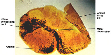
MEDULLA OBLONGATA
Cat, 10% formalin, Marchi stain, 11.5 x.
This is a section of the medulla oblongata, stained by the Marchi* method, which reveals degenerated myelin. Note the degenerated fibers (black) in the pyramid and the crossing of these fibers to form the lateral corticospinal tract. An artifact of tissue preparation is seen in the upper right hand corner of the section. The tissue became folded during handling.
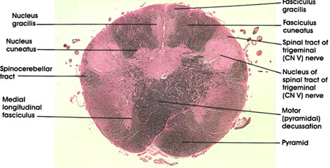
MEDULLA OBLONGATA
Motor (pyramidal) decussation coronal
Human, 10% formalin, Pal-Weigert, 7.5 x.
Nucleus gracilis: One of the dorsal column nuclei. Receives ascending fibers in the dorsal column (fasciculus gracilis) of the spinal cord entering below the level of the sixth thoracic spinal segment. Axons of neurons in this nucleus form the internal arcuate fiber system, cross in the midline (at more rostral levels), and form the medial lemniscus.
Fasciculus gracilis: Dorsal (posterior) column fibers capping the nucleus gracilis. Size is inversely proportional to that of nucleus gracilis, becoming smaller as more fibers terminate in the nucleus.
Nucleus cuneatus: One of the dorsal column nuclei. Located lateral to nucleus gracilis. Receives ascending fibers in the dorsal column of the spinal cord entering above the sixth thoracic spinal segment (fasciculus cuneatus). Axons of neurons in this nucleus, along with axons of the gracile neurons form the internal arcuate fiber system, which crosses in the midline to form the medial lemniscus.
Fasciculus cuneatus: Dorsal (posterior) column fibers capping the nucleus cuneatus. Size is inversely proportional to that of nucleus, becoming smaller as more fibers terminate in nucleus.
Spinal tract of trigerninal (CN V) nerve: Thinly myelinated fibers, hence less densely stained than the heavily myelinated fibers of the spinocerebellar tract. This tract is composed of descending trigeminal fibers and extends from the site of entry of the trigerninal nerve in the pons down to at least the second cervical spinal segment. Synapse in the nucleus of spinal tract of trigerninal nerve. Primarily concerned with pain and thermal sense from the homolateral face.
Nucleus of spinal tract of trigerninal (CN V) nerve: Functionally analogous to and structurally a continuation of the substantia gelatinosa seen at several spinal cord levels. In it terminate fibers of the descending (spinal) tract of cranial nerve V (trigeminal), which enters the neuraxis at a rostral level. The nucleus is primarily concerned with pain and thermal sense from the homolateral face.
Motor (pyramidal) decussation: Constitutes one of the most conspicuous features of sections at this level. Site of crossing of the pyramids to form the lateral corticospinal tracts. Approximately 75 to 90 percent of descending pyramidal fibers cross at this level. The motor decussation forms the basis for voluntary motor control of one half of the body by the contralateral cerebral hemisphere.
Pyramid: Corticospinal fibers prior to decussation.
Medial longitudinal fasciculus: Descending portion of a fiber system with ascending and descending components. Neurons of origin are from various brain stem nuclei with a major vestibular system component. Concerned with movement of neck and head in response to vestibular stimulation.
Spinocerebellar tract: Heavily myelinated. Continuation of the same tracts seen at several spinal cord levels. Destination is the cerebellum
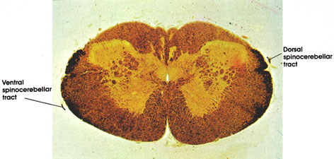
MEDULLA OBLONGATA
Spinocerebellar tract coronal
Dog, Müller's fluid, Marchi's method, 12 x.
This cross section of the lower medulla is stained with the Marchi method, which selectively stains degenerated myelinated tracts black. In this section, black staining outlines the dorsal and ventral spinocerebellar tracts. These are fiber tracts that convey proprioceptive impulses to the cerebellum. They are concerned with unconscious proprioception. Three types of end organs are associated with proprioception: Pacinian corpuscles, muscle spindles, and Golgi tendon organs These receptors detect movements of joints and changes in stretch and tension in muscles and tendons
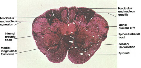
MEDULLA OBLONGATA
Sensory decussation
coronal
Human, 10% formalin, Pal-Weigert and carmine stains, 7 x.
Fasciculus and nucleus gracilis: Note the reduction in size of the fasciculus gracilis as the nucleus gracilis develops. Fibers of the fasciculus synapse on neurons of the nucleus gracilis. The nucleus gracilis appears caudal to the appearance of and terminates caudal to the termination of nucleus cuneatus .
Fasciculus and nucleus cuneatus: Note that the nucleus cuneatus is not as well developed at this level as the nucleus gracilis. The fasciculus cuneatus is voluminous.
Internal arcuate fibers: Second-order fibers arise from gracile and cuneate nuclei, course in the tegmentum of the medulla, and cross in the sensory decussation to form the medial lemniscus. They convey the same modalities of sensation as the gracile and cuneate tracts (proprioception, touch, and vibratory sense).
Medial longitudinal fasciculus: Descending portion of a fiber system with ascending and descending components. Arises from various brain stem nuclei, but with a major vestibular component. This system is concerned with eye and neck movements. The fibers in it are destined to synapse on the motor neurons in the cervical region supplying neck musculature.
Spinal nucleus of V: Continuation of same nucleus seen at more caudal levels .
Spinocerebellar tract: Continuation of the sme tract seen at several caudal levels
Sensory decussation: Also known as decussation of the medial lemniscus. Internal arcuate fibers cross here to form the contralateral medial lemniscus. Provides an anatomical basis for sensory representation of one half of the body in the contralateral cerebral cortex.
Pyramid: Descending corticospinal fibers. Lesion will result in contralateral weakness or paralysis of the upper motor neuron variety.
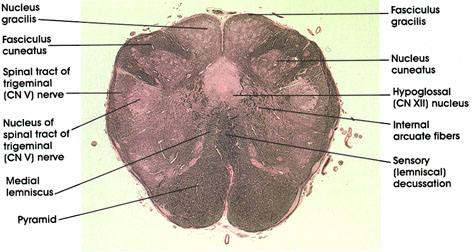
MEDULLA OBLONGATA
Sensory (lemniscal) decussation
coronal
Human, 10% formalin, Pal-Weigert, 5.6 x.
Nucleus gracilis: Fully developed at this level. Only a small remnant of the fasciculus gracilis caps the nucleus.
Nucleus and fasciculus cuneatus: Note that a definite portion of the fasciculus cuneatus caps the nucleus cuneatus compared with that seen in the adjacent nucleus gracilis. The lightly stained island in the fasciculus cuneatus represents neurons of the accessory (lateral) cuneate nucleus.
Spinal tract and nucleus of trigerninal nerve: Continuation of similar structures seen at more caudal levels.
Medial lemniscus: Formed by the decussating internal arcuate fibers. Constitutes the second-order neurons of the posterior column pathways (fasciculi gracilis and cuneatus and their nuclei), conveying kinesthetic sense and discriminative touch to higher levels of the neuraxis.
Pyramid: Corticospinal fibers descending to decussate at more caudal levels.
Sensory (lemniscal) decussation: Also known as decussation of the medial lemniscus. Internal arcuate fibers cross here to form the contralateral medial lemniscus. Provides an anatomical basis for sensory representation of one half of the body in the contralateral cerebral cortex.
Internal arcuate fibers: Second-order fibers arise from gracile and cuneate nuclei, course in the tegmenturn of the medulla, and cross in the sensory decussation to form the medial lemniscus. They convey the same modalities of sensation as the gracile and cuneate tracts (proprioception, touch, and vibratory sense).
Hypoglossal (CN XII) nucleus: A group of large neurons located in a paramedian position dorsal to the medial lemniscus
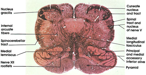
MEDULLA OBLONGATA
Inferior olive
coronal
Human, 10% formalin, Pal-Weigert and carmine stains, 7 x.
Nucleus gracilis: Fully developed at this level. Only a small remnant of the fasciculus gracilis caps the nucleus.
Cuneate nucleus and tract: Note that a definite portion of the fasciculus cuneatus caps the nucleus cuneatus as compared to that seen on the adjacent nucleus gracilis. The lightly stained island in the fasciculus cuneatus represents neurons of the accessory cuneate nucleus.
Spinal tract and nucleus of nerve V: Continuation of similar structures seen at more caudal levels .
Medial longitudinal fasciculus: Note the change of position of the fasciculus in this figure as compared to a more caudal level .This is a result of the formation of the medial lemniscus, which displaces the medial longitudinal fasciculus to a more dorsal location.
Principal and medial accessory inferior olive: This nuclear group distinguishes sections of the medulla at this level. The principal olive is the larger component with its hilum directed medially. The medial accessory olive is found along the border of the medial lemniscus. Inferior olive neurons give rise to olivocerebellar fibers that project into the cerebellum.
Pyramid: See the same structure at more caudal levels.
Internal arcuate fibers: Axons of gracile and cuneate neurons.
Spinocerebellar tract: Continuation of the same tract seen in the spinal cord.
Medial lemniscus: Formed by the decussating internal arcuate fibers. Constitutes the second-order neurons of the posterior column pathways (fasciculi gracilis and cuneatus and their nuclei), conveying kinesthetic sense and discriminative touch to higher levels of the neuraxis.
Nerve XII rootlets: Hypoglossal cranial nerve. Note their characteristic location medial to the inferior olive and lateral to the pyramid. This proximity to the pyramid is the anatomical basis for the inferior or hypoglossal alternating hemiplegia resulting from lesions in this area. This syndrome (also known as medial medullary syndrome) consists of lower motor neuron paralysis of the ipsilateral half of the tongue and contralateral (upper motor neuron) hemiplegia. The hypoglossal nerve supplies all the intrinsic and extrinsic muscles of the tongue except the palatoglossus muscle.
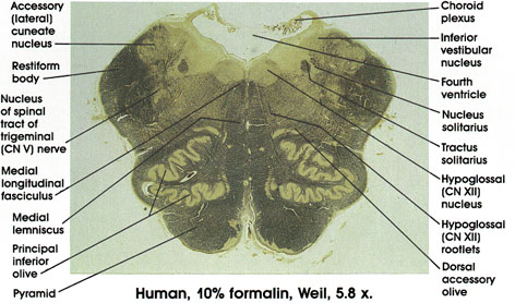
MEDULLA OBLONGATA
Inferior olive
coronal
Choroid plexus: Located in the caudal part of the roof of the fourth ventricle.
Inferior vestibular nucleus: One of four vestibular nuclei. Characteristically located medial and dorsal to the restiform body, and traversed by myelinated bundles.
Tractus solitarius: Contains general visceral as well as special visceral (taste) fibers from the vagus, glossopharyngeal, and facial nerves. Fibers project onto neurons in the nucleus solitarius located in close proximity to the tract.
Nucleus solitarius: Located in close proximity to the tractus solitarius, from which it receives fibers.
Hypoglossal (CN XII) nucleus: A group of large neurons located dorsal to the medial longitudinal fasciculus in the floor of the fourth ventricle in a paramedian position. Rootlets of hypoglossal nerve course in tegmentum of medulla between the medial lemniscus and inferior olive.
Hypoglossal (CN XII) rootlets: Coursing in the tegmentum of the medulla oblongata between the medial lemniscus and the inferior olive. Exit from the ventral surface of the medulla between the pyramid and inferior olive.
Dorsal accessory olive: A component of the inferior olivary complex located dorsal to the principal olive.
Medial longitudinal fasciculus: Descending portion of a fiber system with ascending and descending components. Neurons of origin are from various brain stem nuclei, but with a major vestibular component. The fibers descending in this fasciculus are destined to synapse on motor neurons in the cervical region of the spinal cord, which supply neck musculature.
Accessory cuneate nucleus: Receives fibers of the dorsal spinocerebellar tract entering the spinal cord above the eighth cervical segment. Projects to the cerebellum via the restiform body.
Restiform body: Also known as the inferior cerebellar peduncle. A compact bundle of nerve fibers connecting the medulla with the cerebellum. Described first in 1695 and named by Humphrey Ridley, an English anatomist. Tracts and fibers forming this bundle originate in the medulla and the spinal cord.
Nucleus of spinal tract of trigeminal (CN V) nerve: Receives exteroceptive fibers from the ipsilateral side of the face via the spinal tract of the trigeminal nerve. Lesions result in loss of pain sensation in the ipsilateral face.
Medial lemniscus: Axons of gracile and cuneate nuclei. Forward continuation of the same structure seen in more caudal sections.
Principal inferior olive: Located dorsal and lateral to the pyramid. Note the characteristic convoluted appearance. The principal inferior olive is the largest component of the inferior olivary complex, which includes, in addition, the dorsal accessory inferior olive and the medial accessory inferior olive.
Pyramid: Heavily myelinated motor fiber system. Represents descending fibers from the cerebral cortex that pass through the internal capsule, cerebral peduncle, and pons before reaching the medullary pyramids. Fibers in the pyramid undergo partial crossing in the motor decussation to give rise to the lateral corticospinal tract. It is estimated that, in man, about one million fibers are present in each pyramid.
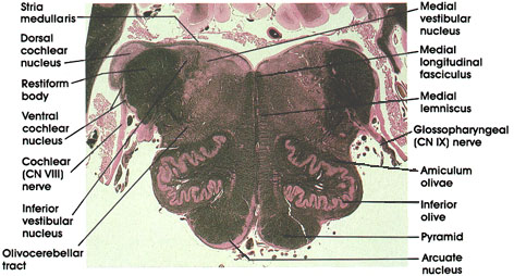
MEDULLA OBLONGATA
Cochleovestibular and
Glossopharyngeal nerves
coronal
Human, 10% formalin, Pal-Weigert, 3.8 x.
Medial longitudinal fasciculus: Descending portion of a fiber system with ascending and descending components. Neurons of origin are from various brain stem nuclei, but with a major vestibular component. This system is concerned with eye and neck movements. The fibers in the descending component are destined to synapse on motor neurons in the cervical spinal cord that supply neck musculature.
Medial lemniscus: Continuation of the same system noted in caudal sections.
Glossopharyngeal (CN IX) nerve: A mixed nerve. Characteristically enters the medulla, inferior and medial to the restiform body.
Amiculurn olivae: A bundle of fibers surrounding the inferior olivary complex. Contains fibers that terminate on neurons of the olivary complex.
Inferior olive: Convoluted laminae of gray matter dorsal to the pyramid. Receives fibers from cortical and subcortical sites and projects fibers primarily to the contralateral but also to the ipsilateral cerebellum via the restiform body. Concerned with motor control.
Pyramid: Heavily myelinated motor fiber system. Contains descending fibers from the cerebral cortex that pass through the internal capsule, cerebral peduncle, and pons before reaching the pyramids. Fibers in the pyramid undergo partial crossing in the motor decussation caudal to this level.
Arcuate nucleus: Motor neurons ventral to the pyramid. Receives cortical input and projects to the cerebellum via the stria medullaris and restiform body. Homologous to pontine nuclei.
Olivocerebellar tract: Axons of neurons in the inferior olivary complex. Fibers arise from both olivary complexes but primarily from the contralateral complex. Destined for the cerebellum via inferior cerebellar peduncle (restiform body). Olivocerebellar fibers constitute the major component of the restiform body.
Cochlear (CN VIII) nerve: Central processes of bipolar neurons in the spiral ganglion. Enters the lateral surface of the pons lateral and dorsal to the restiform body. Projects upon the dorsal and ventral cochlear nuclei. Lesions in the cochlear nerve result in ipsilateral loss of hearing.
Ventral cochlear nucleus: Located ventral and lateral to the restiform body. Receives axons of the cochlear nerve originating in the upper turns of the cochlea.
Restiform body: Also known as inferior cerebellar peduncle. A compact bundle of nerve fibers connecting the medulla with the cerebellum. Tracts and fibers forming this bundle originate in the medulla and the spinal cord.
Dorsal cochlear nucleus: Characteristically located dorsal and lateral to the restiform body. Receives axons of the cochlear nerve originating in the lower turns of the cochlea.
Inferior vestibular nucleus: One of four vestibular nuclei. Characteristically located medial to the restiform body and traversed by myelinated bundles.
Stria medullaris: Axons of arcuate neurons. Courses in the floor of the fourth ventricle. Joins the restiform body to reach the cerebellum.
Medial vestibular nucleus: One of four vestibular nuclei. Characteristically located medial to the inferior vestibular nucleus in the floor of the fourth ventricle. Axons of neurons in this nucleus form the medial vestibulospinal tract.
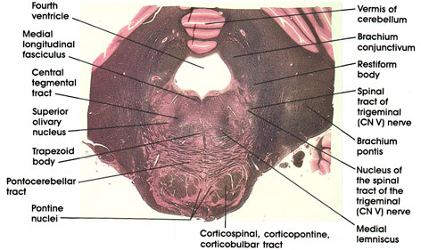
PONS
Trapezoid body coronal
Human, 10% formalin, Pal-Weigert, 2.5 x.
Vermis of the cerebellum: Overlying the fourth ventricle. Midline portion of cerebellum.
Medial longitudinal fasciculus: Continuation of the same structure seen at more caudal levels. Concerned with ocular movement in response to vestibular stimulation.
Central tegmental tract: Compact fiber bundle located dorsal to the medial lemniscus. Carries fibers from midbrain tegmenturn, red nucleus, and periaqueductal gray matter to the inferior olivary complex.
Superior olivary nucleus: One of the tegmental nuclei that belong to the cochlear system. Receives fibers from the trapezoid body and contributes to the formation of the lateral lemniscus.
Trapezoid body: Also known as the inferior acoustic stria. Axons of neurons in the inferior cochlear nucleus form the trapezoid body.
Pontocerebellar tract: Axons of pontine nuclei on their way to the cerebellum via the brachiurn pontis.
Pontine nuclei: Scattered between the descending corticospinal, corticopontine, and corticobulbar fibers and the horizontally oriented pontocerebellar fibers. Receive input from the cerebral cortex via the corticopontine tract and project to cerebellum via the pontocerebellar tract.
Brachium conjunctivum: Also known as the superior cerebellar peduncle. Most important efferent fiber system of the deep cerebellar nuclei. Located dorsolateral to the fourth ventricle. Later in its course, it dips into the tegmenturn of the pons and midbrain .Nerve fibers in this bundle are destined to reach the contralateral red nucleus and ventrolateral nucleus of the thalamus.
Restiform body: Also known as the inferior cerebellar peduncle. Continuation of the same fiber system seen at more caudal levels. The restiform body is shown here entering the cerebellum.
Brachium pontis: A massive bundle of fibers connecting the basal portion of the pons with the cerebellum. Also known as the middle cerebellar peduncle. Contains pontocerebellar tract.
Spinal tract and nucleus of trigerninal nerve: Continuation of the same structures described at caudal levels.
Medial lemniscus: Continuation of the same fiber system described at more caudal levels (see .Note change in orientation of fibers from (previously) vertical in medulla oblongata to horizontal here in the pons.
Corticospinal, corticopontine, corticobulbar tracts: Descending fiber system sectioned transversely. Destined for the pontine nuclei, cranial nerve nuclei, and the spinal cord motoneurons.
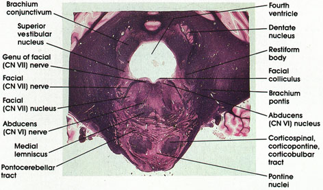
PONS
Facial and abducens
nerves coronal
Human, 10% formalin, Pal-Weigert, 2.2 x.
Genu of facial (CN VII) nerve: A bundle of facial nerve fibers in the floor of the fourth ventricle.
Brachium conjunctivum: Also known as the superior cerebellar peduncle. Most important efferent fiber system of the deep cerebellar nuclei. Located clorsolateral to the fourth ventricle.
Superior vestibular nucleus: Located dorsal and medial to the restiform body. One of four vestibular nuclei. Receives fibers from the vestibular component of the vestibulocochlear (CN VIII) nerve and projects fibers to the cerebellum via the restiform body and to nuclei of extraocular movement via the medial longitudinal fasciculus.
Facial (CN VII) nerve: Coursing ventrolaterally to emerge at the lateral border of the pons.
Facial nucleus: Located medial to the facial nerve. Axons of neurons in the facial nucleus course medially and dorsally to reach the floor of the fourth ventricle (genu of facial nerve) before turning laterally and ventrally to exit from the lateral surface of the pons.
Abducens nerve: Rootlets of the abducens nerve are seen coursing in the tegmenturn of the pons. They arise from the medial aspect of the nucleus and exit from the ventral surface at the caudal border of the pons. Supply the lateral rectus muscle of the eye.
Medial lemniscus: Continuation of the same structure seen at more caudal and more rostral levels.
Pontocerebellar tract: Continuation of the same structure seen at more caudal levels. Axons of pontine nuclei destined for the cerebellum.
Pontine nuclei: Scattered between pontocerebellar fibers and the corticospinal, corticopontine, and corticobulbar fibers. Relay station between the cerebral cortex and cerebellum.
Corticospinal, corticopontine, corticobulbar tracts: Long descending fiber system originating in the cerebral cortex. Sectioned transversely as it passes through the basal portion of the pons.
Abducens nucleus: Located in a paramedian position in the floor of the fourth ventricle. Axons of neurons in this nucleus emerge from the medial aspect of the nucleus to form the abducens nerve. Lesions of the abducens nucleus result in ipsilateral paralysis of lateral gaze. The abducens nucleus and the adjacent genu of facial nerve together form the facial colliculus, a paramedian elevation in the floor of the fourth ventricle.
Brachium pontis: A massive bundle of fibers connecting the basal portion of the pons with the cerebellum. Also known as the middle cerebellar peduncle.
Restiform body: Continuation of the same structure seen at more caudal levels. Seen entering the cerebellum.
Dentate nucleus: The largest of the deep cerebellar nuclei. Axons of this nucleus are major components of the brachium conjunctivum.
Facial colliculus: A paramedian elevation in the floor of the fourth ventricle overlying the abducens nucleus and the genu of the facial nerve
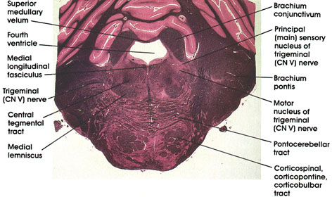
PONS
Trigeminal nerve coronal
Human, 10% formalin, Pal-Weigert, 2.4 x.
Fourth ventricle: The anterior part of the fourth ventricle overlying the pons.
Brachium conjunctivum: Massive outflow tract from the cerebellum seen at this level prior to decussation. Lesions in this area will result in a disorder of coordinated movement. Note the change in position of this structure in more rostral sections .
Principal (main) sensory nucleus of trigerninal (CN V) nerve: Located lateral to the motor nucleus of the trigeminal. Receives touch sensations from the ipsilateral face via the trigeminal nerve.
Motor nucleus of trigerninal (CN V) nerve: Located in the dorsal part of the tegmentum. Axons form the motor root of the trigerninal nerve and supply muscles of mastication, and the tensor tympani, tensor palati, mylohyoid, and the anterior belly of the digastric muscles.
Brachium pontis: Also known as the middle cerebellar peduncle. A massive bundle of fibers connecting the basal portion of the pons with the cerebellum. Contains pontocerebellar fibers.
Pontocerebellar tract: Axons of pontine nuclei destined for the cerebellum via the brachium pontis.
Corticospinal, corticopontine, corticobulbar tracts: Long descending fiber system originating in the cerebral cortex. Sectioned transversely as it passes through the basal portion of the pons. Note the horizontally oriented pontocerebellar tract.
Medial lemniscus: Continuation of the same system seen in more caudal levels.
Trigeminal nerve: Sensory-motor cranial nerve. Seen coursing in the lateral part of the pons.
Central tegmental tract: Compact fiber bundle located in the tegmenturn of the pons. Carries fibers from midbrain tegmentum, red nucleus, and periaqueductal gray matter to the inferior olivary complex.
Medial longitudinal fasciculus: Ascending component of a fiber system originating in vestibular nuclei and destined to synapse with neurons in nuclei of extraocular movement (CN III, IV, and VI). Concerned with control of eye movement.
Superior medullary velum: Forms the anterior (superior) part of the roof of the fourth ventricle.
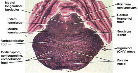
PONS
Trigeminal nerve coronal
Human, 10% formalin, Pal-Weigert, 2.5 x.
Brachium conjunctivum: Also known as superior cerebellar peduncle. Contains axons of deep cerebellar nuclei destined for the red nucleus and thalamus. Forms part of the lateral wall of the fourth ventricle.
Central tegmental tract: A compact fiber bundle located dorsal to the lateral part of the medial lemniscus. Carries fibers from midbrain tegmentum, red nucleus, and periaqueductal gray matter to the inferior olivary complex.
Brachium pontis: Also known as the middle cerebellar peduncle. Massive bundle of fibers connecting the basal portion of the pons with the cerebellum. Contains pontocerebellar fibers from the contralateral half of the pons. Some pontocerebellar fibers from the ipsilateral half of the pons are also contained in the brachium pontis.
Trigeminal (CN V) nerve: A mixed nerve with a larger sensory component (portio major) and a smaller motor component (portio minor).
Pontine nuclei: Located in the basal part of the pons. Continuous caudally with arcuate nuclei in the medulla oblongata. Receive corticofugal fibers and project (pontocerebellar tract) mainly to the contralateral cerebellum.
Corticospinal, corticopontine, corticobulbar tracts: A long descending fiber system sectioned transversely as it courses through the basal part of the pons.
Pontocerebellar tract: Axons of pontine nuclei destined for the cerebellum. Constitutes the major component of the middle cerebellar peduncle (brachium pontis).
Medial lemniscus: Continuation of the same fiber system noted in several more caudal levels. Note the change of orientation of this fiber bundle from a vertical orientation in the medulla to a horizontal orientation at this level.
Lateral lemniscus: Continuation of trapezoid body. Conveys auditory impulses.
Medial longitudinal fasciculus: The ascending component of this fasciculus. Contains fibers from the vestibular nuclei destined for the nuclei of extraocular movement. Lesions of the medial longitudinal fasciculus at this level will result in a characteristic clinical picture known as internuclear ophthalmoplegia
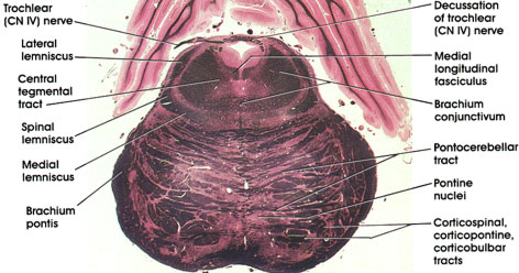
PONS- MESENCEPHALIC JUNCTION
Trochlear nerve coronal
Human, 10% formalin, Pal-Weigert, 2.9 x.
Brachium conjunctivum: Massive outflow tract of the cerebellum. Fibers are seen just prior to and beginning decussation. Fibers project, after decussation, into the red nucleus and ventrolateral nucleus of the thalamus. Lesions in this tract result in a disorder of coordinated movement.
Pontocerebellar tract: The same structure seen at more caudal levels.
Corticospinal, corticopontine, corticobulbar tracts: The same structures seen at more caudal levels. Cut in cross section as they descend to lower caudal levels.
Pontine nuclei: Scattered between pontocerebellar fibers and the corticospinal, corticopontine, and corticobulbar tracts.
Brachium pontis: Axons of pontine nuclei on their way to the cerebellum.
Medial lemniscus: Continuation of the same structure seen at more caudal levels.
Spinal lemniscus: Continuation of the same structure seen at more caudal levels. Contains spinothalamic and spinotectal fibers.
Lateral lemniscus: Contains cochlear fibers. Located laterally and dorsally on its way to the inferior colliculus and medial geniculate body. Concerned with audition.
Trochlear (CN IV) nerve: Seen exiting from the dorsal aspect of the midbrain after decussating. The fourth cranial nerve supplies the superior oblique extraocular muscle. The only cranial nerve to decussate (cross) completely prior to leaving the neuraxis.
Central tegmental tract: Compact fiber bundle located medial to the brachium conjunctivum. Carries fibers from the midbrain tegmentum, red nucleus, and periaqueductal gray matter to the inferior olivary complex. Note change in position of this tract in more caudal levels.
Medial longitudinal fasciculus: Continuation of same structure seen at more rostral and more caudal levels.
Decussation of trochlear (CN IV) nerve: Axons of neurons in the trochlear nucleus seen decussating prior to exit from the dorsal surface of the neuraxis
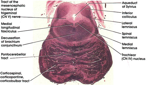
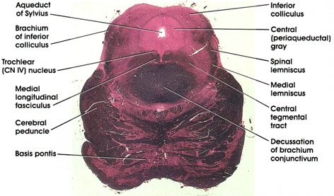
MESENCEPHALON
Inferior colliculus coronal
Human, 10% formalin, Pal-Weigert, 2.7 x.
Aqueduct of Sylvius: Connecting the third and fourth ventricles. Sylvius (Jacques Dubois) was a sixteenth -century French anatomist.
Trochlear (CN IV) nucleus: Motor neurons located in a paramedian position dorsal to the medial longitudinal fasciculus. Axons of neurons in trochlear nucleus decussate prior to leaving the neuraxis).
Inferior colliculus: Ovoid cellular mass in the tecturn of the mesencephalon. Belongs to the auditory system.
Lateral lemniscus: Located laterally and dorsally as it enters the inferior colliculus. Concerned with audition.
Spinal and medial lemnisci: Continuation of the same structures seen at more caudal levels.
Corticospinal, corticopontine, corticobulbar tracts: Sectioned transversely on their way to pontine nuclei, cranial nerve nuclei, and motor neurons of the spinal cord.
Pontocerebellar tract: Axons of pontine nuclei on their way to the cerebellum.
Decussation of brachium conjunctivum: Massive outflow tract of the cerebellum seen decussating at this level. Fibers project, after decussation, into the red nucleus and ventral lateral nucleus of the thalamus.
Tract of the mesencephalic nucleus of trigerninal (CN V) nerve: Processes of pseudounipolar neurons in the mesencephalic nucleus of the trigeminal nerve. Neurons are sparsely scattered on each side of the tract.
Medial longitudinal fasciculus: Continuation of the same structure seen at more rostral and more caudal levels.
MESENCEPHALON
Inferior colliculus coronal
Human, 10% formalin, Pal-Weigert, 3.0 x.
Inferior colliculus: Ovoid cellular mass belonging to the auditory system. Receives fibers from the lateral lemniscus and is reciprocally connected to the medial geniculate body.
Central (periaqueductal) gray: An area of gray matter surrounding the aqueduct of Sylvius. Contains scattered neurons, several nuclei, and some finely myelinated and unmyelinated fibers. Recent interest in this area has focused on its role in pain. The neuropeptide enkephalin has been identified in the central gray.
Spinal lemniscus (spinothalamic and spinotectal tracts): Continuation of the same fiber system seen at more caudal levels.
Medial lemniscus: Continuation of the same fiber system seen at more caudal levels.
Central tegmental tract: A compact fiber bundle located in the dorsal part of the mesencephalon dorsal to the decussation of brachiurn conjunctivurn. Carries fibers from the midbrain tegmentum, red nucleus, and periaqueductal gray matter to the inferior olivary complex. Note how the position of this tract changes in more caudal levels ).
Decussation of brachium conjunctivum: Massive outflow tract of the cerebellum seen crossing in the tegmenturn of the midbrain. Fibers project, after decussation, into the red nucleus and the ventral lateral nucleus of the thalamus. Lesion results in a disorder of coordinated movement.
Basis pontis: Basal part of pons. Contains pontine nuclei as well as corticospinal, corticobulbar, corticopontine, and pontocerebellar fibers.
Cerebral peduncle: Descending corticofugal fiber system. Lesion results in weakness (paresis) or paralysis of the contralateral half of the body, including the face.
Medial longitudinal fasciculus: The ascending component of this bundle. Connects vestibular nuclei with nuclei of extraocular movement (CN III, IV, VI).
Trochlear (CN IV) nucleus: Lies in the V-shaped ventral part of the central gray. Axons arch around the central gray, cross in the anterior medullary velum, and emerge from the dorsal aspect of the mesencephalon. Axons supply the superior oblique extraocular muscle.
Brachium of inferior colliculus: Also known as inferior quadrigeminal brachium. A bundle of nerve fibers from the lateral lemniscus and the inferior colliculus on their way to the medial geniculate body. This fiber bundle conveys auditory impulses from the midbrain to the thalamus.
Aqueduct of Sylvius: Named after the French anatomist Jacobus Sylvius (1478-1555). Connects the third and fourth ventricles.
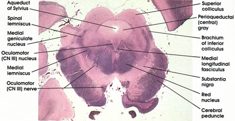
MESENCEPHALON
Superior colliculus coronal
Human, 10% formalin, Pal-Weigert, 2.6 x.
Aqueduct of Sylvius: Connecting the third and fourth ventricles.
Periaqueductal (central) gray: Surrounds the aqueduct of Sylvius. Contains neurons related to both pain inhibition and stimulation.
Superior colliculus: Laminated cellular mass in the tecturn of the mesencephalon. Related to the visual system.
Brachium of inferior colliculus: Also known as inferior quadrigeminal brachium. Bundle of nerve fibers from the lateral lemniscus and the inferior colliculus to the medial geniculate body. Conveys auditory impulses to the thalamus.
Medial longitudinal fasciculus: Continuation of the same structure seen at more rostral and more caudal levels.
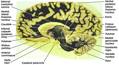
Substantia nigra: Largest nuclear mass in mesencephalon. Sandwiched between the medial lemniscus and the cerebral peduncle. A mass of pigmented cells (melanin) connected with basal ganglia and thalamus. Important in motor control. This area is invariably the site of pathological changes associated with Parkinson's disease. Parkinson was an eighteenth-century English physician.
Cerebral peduncle: Descending corticofugal fiber system. Lesions here result in weakness or paralysis of the contralateral half of the body, including the face.
Red nucleus: So-called because of a pinkish color in the fresh state owing to its high vascularity. This nucleus links the cerebellum, motor cortex, and spinal cord. Major input is from the brachium conjunctivurn and the cerebral cortex. Projects into the motor area of the cortex and spinal cord.
Oculomotor (CN III) nerve: Rootlets of the third cranial nerve seen coursing through the tegmenturn of the midbrain. Note relationship to red nucleus. Supplies the levator palpebrae superioris, the superior rectus, inferior rectus, medial rectus, inferior oblique, and constrictor pupillae muscles. Lesions of the oculomotor nerve result in ipsilateral paralysis of the eye muscles supplied by the nerve as well as a dilated, nonresponsive pupil.
Medial lemniscus: Continuation of the same tract seen at more caudal levels.
Medial geniculate nucleus: A thalamic nucleus concerned with audition. Receives auditory fibers from the inferior quadrigeminal brachiurn (brachiurn of inferior colliculus). Projects to the primary auditory cortex (transverse gyri of Heschl). Heschl was a nineteenth-century Austrian pathologist.
Spinal lemniscus: Continuation of the same structure seen at more caudal levels.
Oculomotor (CN III) nucleus: V-shaped collection of motor neurons located in a paramedian position, dorsal and medial to the medial longitudinal fasciculus. Axons form the oculomotor nerve. The oculomotor nucleus has two components: a somatic motor component, which supplies the extraocular (extrinsic) muscles, and a visceral, parasympathetic component (Edinger-Westphal nucleus), which supplies the intrinsic, constrictor pupillae muscle. Edinger was a nineteenth-century German anatomist and Westphal was a nineteenth-century German neurologist
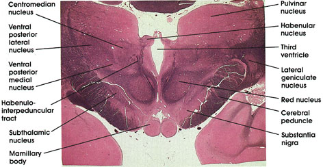
MESENCEPHALON-DIENCEPHALON JUNCTION
coronal
Human, 10% formalin, Pal-Welgert, 2.3 x.
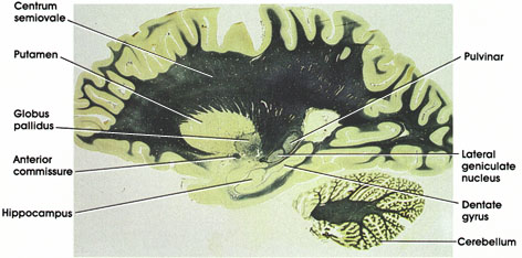
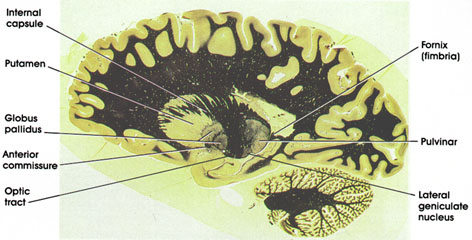
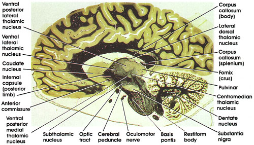
Pulvinar: Belongs to the lateral group of thalamic nuclei. Has reciprocal connections with the medial and lateral geniculate bodies caudally and the association parietal, temporal, and occipital cortices rostrally. Plays a role in several neural functions, including vision, audition, speech, and pain.
Lateral geniculate body: A thalamic relay nucleus concerned with vision. Receives fibers from the optic tract and projects to the primary visual cortex.
Substantia nigra: A mass of pigmented cells containing melanin located dorsal to the cerebral peduncle. This area is invariably the site of pathologic changes associated with Parkinson's disease.
Cerebral peduncle: Descending corticofugal fiber system. Lesion results in contralateral muscle weakness or paralysis.
Oculomotor (CN III) nerve: Coursing in the tegmentum of the midbrain medial to the substantia nigra and cerebral peduncle.
Red nucleus: So-called because of a pinkish color in the fresh state owing to its high vascularity. Links the cerebellum, cerebral cortex, and spinal cord.
Medial geniculate body: A thalamic relay nucleus concerned with audition. Receives fibers from brachium of the inferior colliculus and projects to the primary auditory cortex.
Brachium of superior colliculus: Fiber bundle connecting the superior colliculus and the lateral geniculate nucleus.
Pretectal nucleus: Rostral extension of the superior colliculus. Receives optic tract fibers and projects bilaterally to oculornotor nuclei. Important relay in pupillary light reflex.
Pineal gland: Located dorsal to the mesencephalon. Part of the epithalamus. Has endocrine function and is an important landmark radiologically.
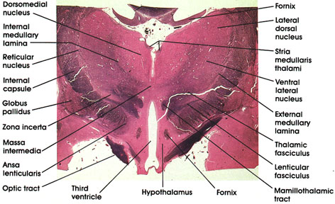
DIENCEPHALON
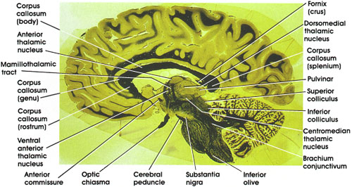
Mamillary body coronal
Human, 10% formalin, Pal-Weigert, 2.4 x.
Pulvinar nucleus: A thalamic nucleus. Belongs to the lateral group of thalamic nuclei. Has reciprocal connections with the medial and lateral geniculate bodies caudally and with the association parietal, temporal, and occipital cortices rostrally. Plays a role in several neural functions, including vision, audition, speech, and pain.
Lateral geniculate nucleus: A thalamic relay nucleus concerned with vision. Receives fibers from the optic tract and projects to the primary visual cortex (calcarine gyrus, area 17).
Red nucleus: So-called because of a pinkish color in the fresh state owing to its high vascularity. Links the cerebellum, cerebral cortex, and spinal cord.
Cerebral peduncle: A massive


 This is a plasma cell with prominent cytoplasmic rough endoplasmic
reticulum. The nucleus has radially arranged clumps of chromatin. The
perinuclear Golgi apparatus does not appear in this plane of sectioning
This is a plasma cell with prominent cytoplasmic rough endoplasmic
reticulum. The nucleus has radially arranged clumps of chromatin. The
perinuclear Golgi apparatus does not appear in this plane of sectioning Plasma cell with prominent cytoplasmic smooth endoplasmic reticulum, normal, electron micrograph
 Platelets, normal, electron micrograph
 Skeletal muscle, normal (inset shows a single sarcomere with dark Z
discs, and a central M line, with thin actin and thick myosin
filaments), electron micrograph
Skeletal muscle, normal (inset shows a single sarcomere with dark Z
discs, and a central M line, with thin actin and thick myosin
filaments), electron micrograph Collagen firbils, normal, electron micrograph
 This electron micrograph of the cilia of the pseudostratified epithelium
of the tracheal mucosa reveals the arrangement of microtubules, with 9
peripheral
This electron micrograph of the cilia of the pseudostratified epithelium
of the tracheal mucosa reveals the arrangement of microtubules, with 9
peripheral  The goblet cells of the gastrointestinal tract are filled with abundant
acid mucin and stain pale blue with this Alcian blue stain.
The goblet cells of the gastrointestinal tract are filled with abundant
acid mucin and stain pale blue with this Alcian blue stain. The cytoplasm of the cells lining this neoplastic gland in a colonic
adenocarcinoma are pink with the mucicarmine stain, indicative of mucin
production typical for an adenocarcinoma at this site
The cytoplasm of the cells lining this neoplastic gland in a colonic
adenocarcinoma are pink with the mucicarmine stain, indicative of mucin
production typical for an adenocarcinoma at this site The areas of black, stippled staining identify the scattered
  Melanin pigment in cells of malignant melanoma, Fontana-Masson stain.
 Lipochrome pigment in the cytoplasm of hepatocytes, H&E stain.
 Hemosiderin, liver, iron stain
 Uric acid crystans, polarized, with red plate
 The red-brown granular material seen here is excessive lysosomal copper
in a patient with the rare autosomal recessive disorder Wilson's
disease. Hepatic copper accumulation results in fatty change (seen here
with cholestasis as well), acute hepatitis, chronic hepatitis, and
eventual cirrhosis
The red-brown granular material seen here is excessive lysosomal copper
in a patient with the rare autosomal recessive disorder Wilson's
disease. Hepatic copper accumulation results in fatty change (seen here
with cholestasis as well), acute hepatitis, chronic hepatitis, and
eventual cirrhosis .
.


 .
.

 .
. 


 .
.


 .
. 


 On the left with H&E staining black mercuric chloride precipitate is
seen in this lymphoma fixed in B-5 and not properly dezenkerized. This
precipitate is seen on the right under polarized light microscopy
On the left with H&E staining black mercuric chloride precipitate is
seen in this lymphoma fixed in B-5 and not properly dezenkerized. This
precipitate is seen on the right under polarized light microscopy


 The lymphocyte is an agranular cell with very clear cytoplasm which stains pale blue. Its nucleus is very large for the size of the cell and stains dark purple. (Notice that the nucleus almost fills the cell leaving a very thin rim of cytoplasm.) This cell is much smaller than the three granulocytes (which are all about the same size). These cells play an important role in our immune response. The T-lymphocytes act against virus infected cells and tumor cells. The B-lymphocytes produce antibodies.