Section 14: Male Reproductive System
The male reproductive organs include the primary sex glands, the testes, the various excretory ducts, the accessory glands, and the penis.
The testes, contained within serous cavities within the scrotum, are compound tubular glands serving both exocrine and endocrine functions. The exocrine function is the formation of the mature male germ cells (the spermatozoa), and the endocrine function is the production of the male sex hormone, testosterone. The testes are ovoid glands approximately 4.5 cm in length and are covered by a thick capsule (tunica albuginea) composed primarily of collagenous connective tissue with some elastic fibers. The tunica albuginea thickens posteriorly to form the mediastinum testis, which is an area where ducts, blood vessels, and nerves leave or enter. From the mediastinum, thin, incomplete, and branching fibrous septa and associated blood vessels radiate into the testis and divide it into about 250 lobules. These incomplete pyramidal spaces contain the seminiferous tubules surrounded by a stroma rich in vascular and cellular elements. Of particular interest in the stroma are the interstitial cells (of Leydig), which secrete the male sex hormone, testosterone. These cells lie within the intertubular or interstitial space, in close proximity to vascular and lymph vascular channels.
The lymphatic endothelium, fibroblasts, and, in many mammalian species, several layers of epithelioid cells possessing many of the fine structural features of smooth muscle ensheath the seminiferous tubules and constitute their tunica propria (boundary) tissue. Apical to the basement membrane lies the seminiferous epithelium, which consists of cells of the germ line that will by means of meiotic divisions and a complex process of cytodifferentiation give rise to the haploid male gamete, the spermatozoon. The production of sperm by the epithelium proceeds in an orderly, cyclic fashion from the base of the epithelium' to its apex, as well as in a coordinated, wave-like manner along the length of the tubule. The mitotic and meiotic divisions in the more basal aspects of the epithelium displace the developing sex cells progressively farther away from the basement membrane, with the more mature sex cells occupying, therefore, the most apical (luminal) position.
Cytoplasmic processes of a second population of cells, termed Sertoli cells (also sustentacular or nurse cells), extend from the basement membrane to the most apical portion of the seminiferous epithelium. Sertoli cells are somatic cells that establish an intimate morphological and physiological relationship with each of the developing germ cells, from the earliest, most primitive gonial cell at the basal lamina, to the apically situated mature spermatids ready to be shed into the tubular lumen. Because of the cyclic nature of the process of spermatogenesis occurring in all seminiferous tubules and the microtome knife's random entry into this cycle, the student must be reconciled to studying many sections of seminiferous tubules in order to appreciate and correctly identify the named germ cells of the epithelium and their place in the process of spermatogenesis.
As in other renewing epithelia with which the student is already familiar, the most primitive, regenerative cells lie at the basement membrane of the epithelium. In the testis, these cells are termed the spermatogonia (and are diploid). They divide mitotically and renew the gonial stem cell population. However, a subpopulation of spermatogonia is programmed to enter meiotic prophase and thus become primary spermatocytes. As daughter cells of spermatogonia, the primary spermatocytes reside just apical to the layer of spermatogonia. These cells, the largest germ cells, are recognized by their distinct meiotic nucleus, as well as by their position near the spermatogonia at the base of the epithelium. Primary spermatocytes undergo first meiotic division, giving rise to secondary spermatocytes; these cells occupy a place within the seminiferous epithelium just apical to the primary spermatocytes. The secondaries are short-lived (and thus not frequently observed within sections of the germinal epithelium); secondaries complete second meiotic division and give rise to round spermatids, which occupy an adluminal site within the epithelium. A complex process of cytodifferentiation ensues, whereby these round cells are transformed into elongate spermatids and, finally, spermatozoa.
There exists no "free space" within the seminiferous epithelium, all interstices being filled with Sertoli cell cytoplasm. Because of the attenuated nature of the Sertoli cell processes within the seminiferous epithelium, it is not possible to distinguish cell-to-cell boundaries between Sertoli cells. For this reason, the Sertoli cells were considered prior to the advent of electron microscopy to be syncytial. Indeed, the occluding tight junctions between Sertoli cells are now recognized as the morphological basis for the "blood-testis" (blood epithelial) barrier. Because of the placement of the tight junctions just apical to the gonial stem cells, the seminiferous epithelium is segregated into a basal compartment, open to the interstitium, and an apical compartment containing the meiotic cell line (considered non-self by the body's immune surveillance system). Thus, the haploid spermatids develop within an immunologically privileged compartment maintained by the Sertoli-Sertoli cell occluding junctions.
Within the seminiferous tubules (also termed tubuli contorti in recognition of the convoluted course that they describe with in lobules), sperm are transported along their lengths by means of a remarkably high fluid flux across the epithelium. Upon reaching the epididymis, the spermatozoa undergo additional physiological maturation (capacitation) and gain fertilizing capacity and motility; there, also, the testicular fluid is resorbed.
Leading from the seminiferous tubules are the tubuli recti or straight tubules lined only with columnar cells apparently derived from Sertoli cells. The straight tubules open into the rete testis, a network of irregular, anastomosing channels lined with a simple cuboidal or columnar epithelium, which may possess a single flagellum. The efferent ductules (10 to 15 in man) emerge from the rete testis and join to form a portion of the head of the epididymis. Histologically, each efferent duct presents a stellate luminal profile, reflecting an epithelium in which ciliated columnar cells alternate with non-ciliated cuboidal cells. The epididymal duct in man is a highly coiled tube about 5 m in length. It is lined with a pseudostratified columnar epithelium with tall columnar cells and rounded basal cells. The columnar cells possess long non-motile stereocilia. The ductus deferens is continuous with the epididymis. The lumen of the ductus deferens increases in size, and the wall thickens as it extends distally. Near the prostate gland, the ductus is enlarged to form the ampulla; immediately thereafter, it is joined by the duct from the seminal vesicle. The duct formed by this confluens, the ejaculatory duct, courses through the substance of the prostate gland and empties into the prostatic urethra. It is lined with a pseudostratified or simple columnar epithelium.
Three major accessory sex glands are associated with the male reproductive system: the seminal vesicles, the prostate gland, and the bulbourethral (Cowper's) glands. The seminal vesicles develop as outgrowths of the ductus deferens. Each is a glandular sac honey-combed by thin branching folds of the mucosa lined with a pseudostratified columnar or cuboidal epithelium. The prostate gland encircles the urethra adjacent to the neck of the bladder and is formed of 30 to 50 tubuloalveolar glands grouped into lobes. The glandular epithelium consists of simple cuboidal or columnar cells. Prostatic concretions (corpora amylacea) are prominent constituents of the alveoli. The bulbourethral glands are compound tubuloalveolar glands that secrete a clear, viscous mucoid product. The secretory epithelium is cuboidal to columnar. The ducts of the gland enter the cavernous urethra. The secretions of each of these glands are important physiological components of semen.
The penis serves as an outlet for urine and semen and as a copulatory organ. The penis is made up of three cylinders of erectile tissue. These include the corpus spongiosum, which enlarges distally into the glans penis; parallel with, and dorsal to, the corpus spongiosum are the paired corpora cavernosa, which extend distally to the glans. The corpora cavernosa are united distally by a median partition, the pectiniform septum. All three structures are surrounded by a thick fibrous tunica albuginea and a subcutaneous connective tissue layer covered by a thin skin.
Some names associated with the male reproductive system follow: Cowper was a seventeenthcentury English anatomist, von Leydig, a nineteenth-century German anatomist, and Sertoli, a nineteenthcentury Italian histologist.
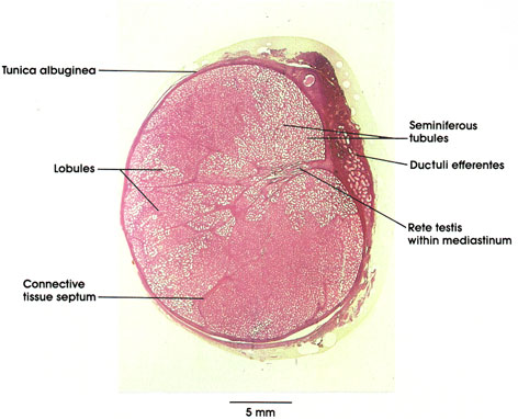
The integrity of the delicate, tubular parenchyma of the male gonad is maintained by the robust connective tissue capsule of white fibrous connective tissue, the tunica albuginea, and the septa, which project interiorly, dividing the organ into lobules. The lobules contain the seminiferous tubules, lined with the specialized seminiferous epithelium, which gives rise to the male gametes, the spermatozoa. In addition, a posterior connective tissue mass (the mediastinum) projects to the interior of the organ and provides an avenue for the egress of fluid and spermatozoa from the seminiferous tubules. Irregular, epithelium-lined, anastomosing channels form a network, the rete, within the mediastinum. From these channels, the ductuli efferentes arise, pierce the tunica albuginea, and subsequently form, in man, the first portion of the epididymis.
Neither the seminiferous tubules nor the intratesticular duct system have significant muscular coats, or motile cilia by which to move the tubular contents. This underscores the role of fluid flux in moving the spermatozoa through the initial portion of the excurrent duct system. The ductuli efferentes are the sole portion of the male duct system that bears motile cilia on the lining epithelial cells.
The testis is invested on its anterior and lateral aspects by a serous envelope, the tunica vaginalis, which is a peritoneal vestige carried with the testis in its descent to the scrotum. The testis is cushioned by and glides within this serous envelope, relieving compression, to which the organ is exquisitely sensitive.
Periphery
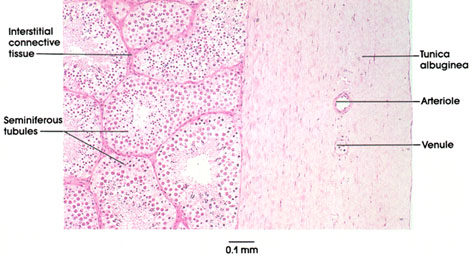
The testis is enclosed by a robust dense white fibrous connective tissue capsule, the tunica albuginea, designated for its glistening white appearance in the unfixed state. Vascular elements pierce the tunica albuginea and travel within septa that are continuous with the intertubular connective tissue of the organ; posteriorly, the tunica albuginea is specialized as the mediastinum testis. Deep to the tunica, note the many profiles of seminiferous tubules, which are lined with the complex seminiferous epithelium. The epithelium is seen to better advantage in later plates at higher magnification.
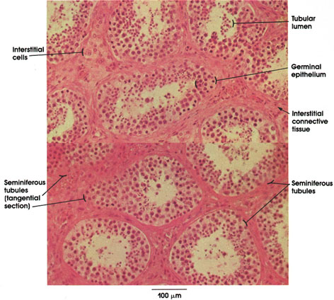
This is a section of the testis showing the seminiferous tubules separated by interstitial connective tissue. The seminiferous tubules are lined by a stratified epithelium, the germinal epithelium, composed primarily of sex cells with some supporting cells. The epithelium rests on a basement membrane that varies in thickness with age. The lumina of the seminiferous tubules contain mature sex cells (spermatozoa). Seminiferous tubules are separated by an interstitial stroma made up of loose connective tissue and containing the interstitial cells of Leydig. These large ovoid cells that occur in groups secrete testosterone, the male sex hormone.
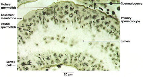
In the seminiferous tubule, spermatogenic cells are arranged in orderly layers between the basement membrane and the lumen.
Spermatogonia: Located directly above the basement membrane. Spherical nucleus. Spermatogonia are the germ cells from which spermatozoa ultimately arise. They are the only sex cells present before onset of puberty. They contain 23 pairs of chromosomes.
Primary spermatocytes: Lie in the next layer, deep to the spermatogonia. Largest germ cells. Nuclei are large and vesicular with condensed chromatin. Chromatin may appear as elongated spiremes, i.e., irregularly disposed chromatin filaments. Primary spermatocytes divide by meiosis. Meiosis is nuclear division in which the diploid chromosome number (23 pairs) is halved to the haploid number (23 single set) in the formation of sex cells.
Round spermatids: Adjacent to the lumen. Small in size. Spermatids constitute the last stage in the transformation to spermatozoa.
Mature spermatids: Heads are located near Sertoli cells, and tails project into the lumen. Heads are transformed nuclei of round spermatids. Will be released into lumen as spermatozoa.
Sertoli cells: Supporting cells of the testicular epithelium, which were first described by the Italian physiologist Enrico Sertoli in 1865. Tall columnar cells extend from the basement membrane to the lumen. Nucleus ovoid in shape with prominent nucleolus. Cell borders are difficult to outline in this preparation.
The process of spermatogenesis from spermatogonia to mature spermatozoa requires about 64 days in man.
Seminiferous epithelium
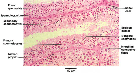
Plastic-embedded, 1.5 µm, sections reveal to particular advantage the cell types constituting the seminiferous epithelium. The longitudinal section of a tubule in the center of the field contains many such representative cell types. Note the spermatogonia on the basement membrane. The largest of the germ cells, the primary spermatocytes with their characteristic meiotic prophase nuclei, occupy the region of the epithelium just apical to the basal layer of spermatogonia. The infrequently seen secondary spermatocytes are intermediate in size and placement within the epithelium between primary spermatocytes and spermatids.
Most apically situated within the epithelium are the spermatids, which possess either the smallest round nuclei of any cell of the epithelium or elongate, condensed nuclei, depending upon their stage of maturation toward mature spermatozoa.
The eosinophilic cytoplasmic remnants resulting from the differentiation of round spermatids to elongate spermatids are seen at the luminal surface and are termed residual bodies. Sustentacular cells, the Sertoli cells, phagocytize much of this residual cytoplasm. The nuclei of Sertoli cells are identified by their distinctive urn shape and prominent nucleolus. Note also how clusters of spermatids develop synchronously, enveloped within the apical extensions of Sertoli cell cytoplasm.
Adjacent seminiferous tubules share a common lamina propria; an area of interstitial connective tissue is shown where three adjacent tubules meet. Leydig cells are commonly found in such areas but are not present in this particular section.
Seminiferous epithelium
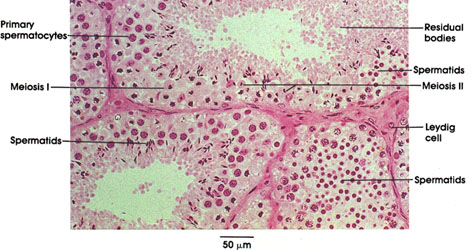
The process of spermatogenesis is best appreciated from the study of the various stages represented in adjacent sections of a seminiferous tubule. Occasionally, however, a tubule is cut in fortuitous tangential section in such a manner that the processes occurring along its length can be appreciated. Such is the case in this figure, in which, toward the upper left of the section, primary spermatocytes are seen; these are the daughter cells of the basally situated spermatogonia. Note their distinctive meiotic prophase nuclei. Division of primary spermatocytes (meiosis 1) gives rise to daughter cells, the secondary spermatocytes. These, in turn, complete the second meiotic division, giving rise to haploid daughter cells, the round spermatids (upper right). Over time, the round spermatids complete cytodifferentiation and become mature, elongate spermatids. These cells lie most apically within the epithelium, among the cytoplasmic residual bodies. Mature spermatids shed into the tubular lumen are termed spermatozoa. Other tubules (such as the one in the lower right portion of the figure) may be cross-sectioned tangentially such that the plane of section passes predominantly through one cell population of the seminiferous epithelium. This accounts for the absence of a luminal profile in this tubule and the predominance of round spermatids.
Testis
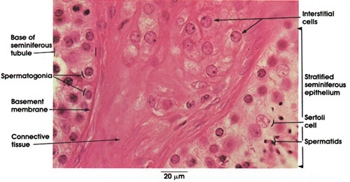
This figure shows parts of two seminiferous tubules separated by a connective tissue sheath. Within this connective tissue sheath are embedded large ovoid cells, the interstitial cells of Leydig. These occur in groups, have a rounded, large eccentric nucleus with a prominent nucleolus and a vacuolated cytoplasm, which results from the loss of lipid droplets and crystals during tissue processing. The interstitial cells are the source of testosterone, the male sex hormone, whose functions include the development and maintenance of secondary sex characteristics and the structure and function of the male accessory organs, the development of psychosexual behavior (in part) in the mature male, a role in protein metabolism, and the regulation of the output of the pituitary gonadotropic hormone, interstitial cell-stimulating hormone (ICSH). The seminiferous tubules shown reveal part of their contents, spermatogonia, spermatids, and Sertoli cells
Straight tubules and rete testis
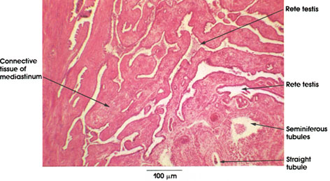
The seminiferous tubules of the testis (tubuli contorti) open into the straight tubules (tubuli recti). The latter are short, straight tubules lined with a single layer of tall columnar Sertoli cells. The straight tubules empty into a system of irregular, anastomosing epithelium-lined cavernous spaces, the rete testis, located in the dense connective tissue of the mediastinum.
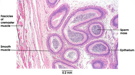
The epididymis, a highly coiled duct approximately 6 m in length in man, is firmly adherent to the gonad. in histological preparations, therefore, it is not unusual to observe sections of both testis and epididymis. The duct is divided anatomically into three major portions (head, body, and tail) and by histological criteria, several further subdivisions may be recognized. Because of the tight coiling of the duct, histological sections invariably reveal many tubular profiles in different planes of section.
The portion of the epididymis shown here is from the distal portion of the head, in which the epithelial lining of the duct is a tall pseudostratified columnar epithelium, lacking goblet cells. The tall columnar cells bear prominent (non-motile) stereocilia on their apical surfaces, facilitating the resorption of testicular tubular fluid in which the spermatozoa are transported to the organ. A thin coat of smooth muscle surrounds the duct and is readily differentiated from the loose connective tissue surrounding the coils of the duct. A packed mass of non-motile sperm is seen within the lumen of the epididymis. In this preparation, striated muscle fibers belonging to the cremaster muscle are seen investing the organ. This muscle of the spermatic cord enables the gonad to be retracted within the scrotum toward the, abdomen.
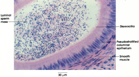
the tall pseudostratified columnar epithelium is demonstrated to advantage. The luminal cells bear remarkable numbers of highly developed stereocilia on their apical surfaces. The lumen of the duct contains a packed mass of spermatozoa. Sperm gain motility and fertilizing capacity during their passage through the epididymis.
Cross section
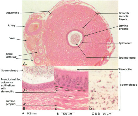
A. 50 x., B. 162 x., C. & D. 612 x.
The ductus deferens (A) is the most prominent component of the spermatidicord and is readily palpated through the scrotal skin. Within the cord, the vas lies in company with a plexus of veins, the pampiniform plexus (so-named for the resemblance of the tortuous veins to tendrils of a vine), branches of the spermatic artery and nerves of the spermatic plexus, and the cremaster muscle.
The mucosa of the vas deferens consists of a prominent pseudostratified columnar epithelium bearing conspicuous stereocilia (B and C and is underlaid by a lamina propria, which abuts the robust muscular components of the organ without a defined submucosal layer. The musculature of the organ is organized into sparse inner longitudinal, prominent middle circular, and well-defined outer longitudinal layers. The vas is surrounded by an adventitia throughout its length. The organ's overall topography, together with the tendency of its mucosa to be thrown into deep folds upon fixation, causes this organ to be readily mistaken for other muscular ducts of the body (i.e., ureter and oviduct). Careful examination of the lining epithelium will confirm identification of each; in addition, the vas may exhibit a packed mass of luminal sperm (D). At the time of ejaculation, the vas rhythmically contracts, conveying spermatozoa stored within the terminal portion of the epididymis and along the length of the vas itself to the ejaculatory ducts. Electron-microscopic studies of the vas have confirmed that its fine structure differs significantly along its length and that the description of the vas as a passive conduit for sperm is inadequate. The scrotal ductus deferens (frequently referred to clinically as the vas) is the common site of ligation or excision of a portion of the organ in the surgical procedure of vasectomy.
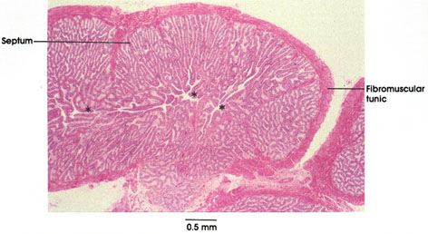
The seminal vesicles arise as tortuous diverticula of the vas deferens and therefore present histologically as multiple sections through a tubular structure. The wall consists of adventitia, muscularis, and elaborate mucosa, thrown into several orders of branching folds. The folds, like villi of the intestinal tract, are underlaid by a tunica propria of loose connective tissue. Secretions produced by the epithelial cells are delivered to a central luminal channel*, the extent of which is largely obscured by the tortuosity of the tube. Lobulation of the organ by septa from the adventitia is incomplete and inconspicuous
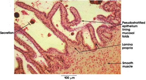
The seminal vesicle is a diverticulum of the adjacent ductus deferens with a remarkably folded mucosal lining of pseudostratified cuboidal or columnar epithelium projecting into the lumen. The lumen frequently contains acidophilic rounded secretion masses. Underlying the epithelial lining is a thin supporting sheath of connective tissue, the lamina propria, which extends into the mucosal folds. Beneath the lamina propria is a coat of smooth muscle fibers consisting of an inner circular layer and an outer longitudinal layer.
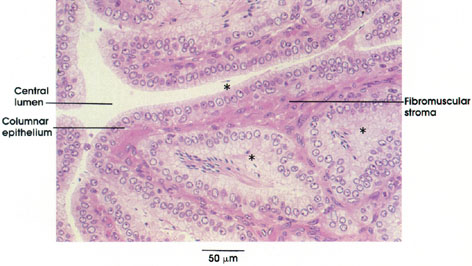
The complex ramifications of the mucosa are shown; the folds are lined by a columnar to pseudostratified columnar epithelium. Epithelial development is androgen-dependent. The height of the epithelium in this specimen suggests ample hormonal stimulation and active secretion. Post-mortem migration of spermatozoa (asterisks) into the recesses and pockets formed within the mucosal lining gave rise to the earlier erroneous interpretation that the seminal vesicle serves as a reservoir or storage depot for sperm. The gland is in fact a major secretory accessory sex gland, the secretions of which, in man, contribute the majority of the seminal volume.
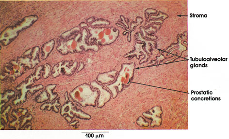
Stroma: Abundant and continuous with the gland capsule, it constitutes one third to one fourth of the gland volume and is composed of fibroelastic connective tissue intermixed with smooth muscle fibers. Glands are embedded in the stroma.
Tubuloalveolar glands: Irregular, large lumen, widely spaced tubules with alveolar extensions, which vary greatly in shape and size. Epithelial lining in tissue sections is simple cuboidal to columnar in shape, depending upon physiological state.
Prostatic concretions: Corpora amylacea, acidophilic condensed secretions of prostatic glands. They may be lamellated and increase in number with advancing age. Source of prostatic calculi.
The prostate is located at the origin of the urethra (which it surrounds), adjacent to the urinary bladder. The prostate secretes a thin, opalescent, slightly acid fluid, which contains several enzymes, including diastase and proteases, and citric acid. The smaller prostatic concretions are found in the prostatic fluid.
Cross section
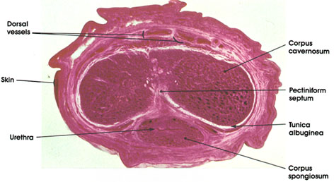
The penis is formed primarily of three cylindrical masses of erectile tissue. Note the paired corpora cavernosa and the ventrally placed corpus spongiosum (corpus spongiosum urethrae) containing the urethra. A dense collagenous tissue capsule, the tunica albuginea, surrounds the corpora cavernosa. This capsule fuses in the midline to form the pectinate septum, which is thickest and most complete near the root of the penis. The tunica albuginea of the corpus spongiosum is thin. Each corpus consists of a network of cavernous vascular sinuses lined with endothelium, separated by fibromuscular trabeculae composed of connective tissue and smooth muscle fibers.
The three corpora are encompassed by a common, loose connective tissue fascia rich in elastic fibers and a thin skin. Note the dorsal vessels (arteries and veins) of the penis, located in the fascia, which are part of the complicated blood supply of this organ.
Corpus cavernosum
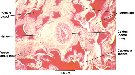
The erectile tissue of the corpus cavernosum of the penis is composed of cavernous spaces separated by fibromuscular septa or trabeculae. The latter are extensions of the tunica albuginea, the fibrous coat that surrounds the corpus. The cavernous spaces are filled with blood, and the engorgement of these spaces results in the erection of the penis. Note the central (deep) artery, which traverses the corpus cavernosum. This artery gives rise to the spiraling helicine arterioles that open into the sinuses. The central artery is the principal vessel for filling the sinuses during erection.
Adjacent to the central artery, note the nerve cut in cross section. The penis is richly supplied with spinal, sympathetic, and parasympathetic fibers. The autonomic fibers innervate the smooth muscle in the arterial wall and trabeculae.