Section 17: Central Nervous System
Ventral lateral nucleus coronal
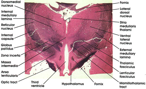
Fornix: A paired C-shaped fiber tract connecting the hippocampus with several brain regions, including the mamillary body, anterior thalamic nucleus, septal nuclei, and cingulate gyrus. Seen in two locations in this figure, below the corpus callosurn and within the hypothalamus as it approaches its termination in the mamillary body.
Massa intermedia: Connects the two thalami across the third ventricle. Present in about 70 percent of human brains. Also referred to as the interthalamic adhesion.
Lateral dorsal (thalamic) nucleus: Belongs to the dorsal subgroup of the lateral thalamic nuclei. Has an association function.
Ventral lateral (thalamic) nucleus: Traversed by bundles of myelinated fibers. Located lateral to the internal medullary lamina. Receives fibers from the cerebellum via the brachium conjunctivum (dentatorubrothalamic fiber system). Has reciprocal connections with the primary motor cortex. Plays a role in motor control.
External medullary lamina: Defines the lateral boundary of the thalamus.
Thalamic fasciculus: A bundle of myelinated fibers destined for several thalamic nuclei from the basal ganglia and cerebellum.
Lenticular fasciculus: A bundle of myelinated fibers originating from the basal ganglia (globus pallidus) and destined for the thalamus via the thalamic fasciculus.
Mamillothalamic tract: Seen in cross section. Connects the mamillary body with the anterior thalamic nucleus.
Hypothalamus: Ventral to the thalamus. Forms part of the lateral wall of the third ventricle. The fornix separates medial and lateral zones of the hypothalamus.
Third ventricle: Between the two hypothalami and two thalami. Bisected by the massa intermedia.
Optic tract: Myelinated fiber bundle conveying visual impulses from the contralateral field of vision to the lateral geniculate nucleus.
Ansa lenticularis: Axons of neurons in globus pallidus coursing around the internal capsule. Projects to ventral anterior thalamic nucleus. Together with fibers from lenticular fasciculus forms the thalamic fasciculus.
Zona incerta: Sandwiched between the lenticular fasciculus and thalamic fasciculus. Rostral continuation of mesencephalic reticular formation. Continuous with the reticular nucleus of the thalamus.
Globus pallidus: Belongs to the basal ganglia along with the caudate nucleus and putamen. Located medial to the putamen and traversed by heavily myelinated fiber bundles. Comprises the principal efferent nucleus of the basal ganglia. Receives fibers from caudate, putamen, and subthalamic nuclei; and projects fibers to the thalamus (ventral anterior nucleus) and the subthalamic nucleus.
Internal capsule: The posterior limb of the internal capsule separates the basal ganglia and the thalamus. Massive cortical afferent and efferent bundle.
Internal medullary lamina: A band of myelinated fibers that separate the medial from the lateral group of thalamic nuclei. Contains fibers that interconnect different thalamic nuclei.
Reticular nucleus: A thalamic nucleus located between the internal capsule and external medullary lamina.
Dorsomedial (thalamic) nucleus: Belongs to the medial group of thalamic nuclei. Most highly developed in man. Located medial to the internal medullary lamina. Has reciprocal connections with the prefrontal cortex and hypothalamus. Receives input from other thalamic nuclei. Concerned with affective behavior and memory.
Stria medullaris thalami: Located clorsomedial to the thalamus. A component of the epithalamus. Connects the septal area and the habenular nuclei.
Anterior commissure coronal
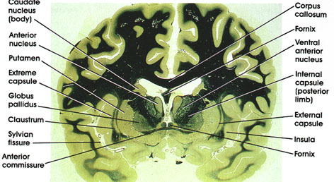
Corpus callosum: A massive bundle of myelinated fibers connecting the two hemispheres. Important in interhemispheric transfer of information.
Fornix: C-shaped, paired fiber system connecting the hippocampus with several brain regions, including the mamillary body, anterior thalamic nucleus, septal nuclei, and cingulate gyrus. Seen in two locations in this figure, beneath the corpus callosurn and above the anterior commissure.
Ventral anterior (thalamic) nucleus: One of the lateral group of thalamic nuclei. Characteristically traversed by heavily myelinated fiber bundles. Receives fibers from the basal ganglia and is reciprocally connected with the cerebral cortex. Plays a role in motor control.
Internal capsule (posterior limb): Separates the thalamus from the basal ganglia (putamen and globus pallidus). Carries fibers from and to the cerebral cortex. Lesions result in contralateral motor and sensory deficits.
External capsule: An efferent cortical bundle sandwiched between the putamen and claustrum.
Insula: Also referred to as the island of Reil,* lies deep in the sylvian fissure. Concerned primarily with autonomic function.
Caudate nucleus (body): A component of the basal ganglia. The body of the caudate is continuous rostrally with the head of the caudate and caudally with its tail. Plays a role in motor control.
Anterior (thalamic) nucleus: Belongs to the anterior group of thalamic nuclei. Has reciprocal connections with the mamillary body via the mamillothalamic tract and with the cingulate gyrus via the internal capsule. Considered part of the limbic system and thus plays a role in emotional behavior and memory.
Putamen: One of the basal ganglia nuclei. Concerned with motor control.
Extreme capsule: An efferent cortical bundle. Situated between the claustrum and the insula.
Globus pallidus: One of the basal ganglia nuclei. Located medial to putamen and characteristically traversed by heavily myelinated fiber bundles. Receives fibers from the caudate and putamen and projects to the thalamus (ventral anterior nucleus). Has also reciprocal connections with the subthalamic nucleus.
Claustrum: A thin layer of gray substance located between the external and extreme capsules.
Sylvian fissure: A major fissure on the lateral surface of the hemisphere separating the temporal from the frontal and parietal lobes.
Anterior commissure: A compact fiber bundle in close proximity to the fornix. Interconnects the olfactory bulbs and the temporal cortices.
*Reil was an eighteenth -century German physician, neurologist, and histologist.
Ventral anterior nucleus coronal
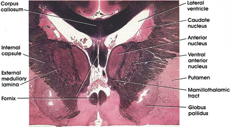
Corpus callosum: Massive fiber bundle connecting the two cerebral hemispheres. Important in interhemispheric transfer of information.
Caudate nucleus: Basal ganglia nucleus. Important in motor control.
Anterior (thalamic) nucleus: Located in rostral diencephalon. Receives the mamillothalarnic tract from the mamillary body and projects to the cingulate gyrus of the cerebral cortex. A component of the limbic system.
Ventral anterior (thalamic) nucleus: Medial to the internal capsule. Traversed by heavily myelinated fiber bundles. Receives fibers from the basal ganglia and is reciprocally connected with the motor cortex. Plays a role in motor control.
Putamen: One of the basal ganglia nuclei. Similar to the caudate nucleus in structure and connections. Along with the caudate it forms the neostriaturn. Lies ventral and lateral to the internal capsule.
Globus pallidus: Another of the basal ganglia nuclei. Receives fibers from caudate and putamen and projects to the thalamus (ventral anterior nucleus). Also reciprocally connected with the subthalamic nucleus.
Mamillothalamic tract: Seen entering the anterior nucleus of the thalamus. Axons of neurons in the mamillary body.
External medullary lamina: Between the internal capsule and ventral anterior nucleus. Defines the lateral boundary of the thalamus.
Internal capsule: The posterior limb, separates the basal ganglia (putamen and globus pallidus) from the thalamus.
Fornix: At this rostral level, the fornix bundle separates as the columns of the fornix arch ventrally and caudally en route to the mamillary body
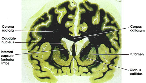
Corpus callosum: A massive myelinated fiber bundle connecting the two hemispheres. Important in interhemispheric transfer of information.
Putamen: Located lateral to the anterior limb of the internal capsule. Part of the basal ganglia (along with the caudate nucleus and globus pallidus). Concerned with regulation of movement.
Globus pallidus: Belongs to the basal ganglia (along with caudate nucleus and putamen). Located medial to putamen, and traversed by heavily myelinated fiber bundles. Constitutes the principal efferent nucleus of the basal ganglia. Receives fibers from the caudate, putamen, and subthalamic nuclei and projects fibers to the thalamus (ventral anterior nucleus) and to the subthalamic nucleus.
Corona radiata: White matter core of the cerebral hemispheres. Contains afferent and efferent fibers to and from the cerebral cortex.
Caudate nucleus: C-shaped mass of gray matter closely related to the lateral ventricle. Component of the basal ganglia. Plays a role in motor control.
Internal capsule (anterior limb): Separates the caudate nucleus and putamen. Carries fibers from and to the cerebral cortex
Neostriatum (caudate nucleus and putamen)
coronal
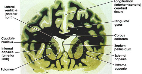
Longitudinal (interhemispheric) cerebral fissure: Separates the two cerebral hemispheres. Filled with a dural fold, the falx cerebri, which has been stripped away in this preparation.
Cingulate gyrus: Located dorsal to the corpus callosum. A component of the limbic lobe, which, in addition to the cingulate gyrus, includes the subcallosal gyrus, isthmus, parahippocampal gyrus, and uncus.
Corpus callosum: A massive myelinated fiber bundle connecting the two hemispheres. important in interhemispheric transfer of information.
Septum pellucidum: A thin septum, ventral to the corpus callosum, separates the two lateral ventricles. May contain a cavity, the cavurn septi pellucidi.
External capsule: One of the efferent cortical bundles. The others include the internal and extreme capsules. Located lateral to the putamen.
Extreme capsule: One of the efferent cortical bundles. Lateral to the external capsule. Between the external and extreme capsules is the claustrum (not seen in this preparation).
Lateral ventricle: Anterior horn of the lateral ventricle. Note the characteristic bulging of the head of the caudate nucleus into the cavity of the ventricle.
Caudate nucleus: C-shaped mass of gray matter closely related to the lateral ventricle. The part seen in this figure is the head of the caudate, which has a characteristic bulge into the lateral ventricle. This characteristic bulge is lost in the disease Huntington's chorea. The caudate nucleus is part of the basal ganglia, and thus plays a role in the regulation of movement. Huntington was a nineteenth-century American physician.
Internal capsule: Heavily myelinated broad band of white substance that arises from wide areas of the cerebral cortex and descends to the brain stem and spinal cord. Carries fibers from and to the cerebral cortex. The anterior limb of the internal capsule separates the caudate nucleus and putamen.
Putamen: Located lateral to the anterior limb of the internal capsule and medial to the external capsule. The putamen, like the caudate, is a component of the basal ganglia and thus plays a role in motor control.
Deep cerebellar nuclei
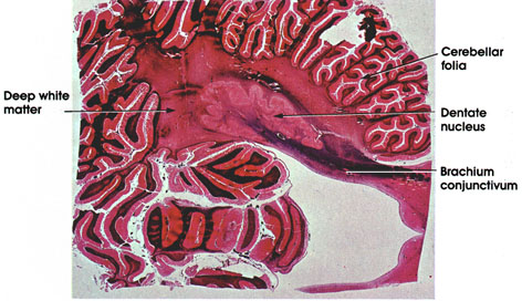
Cerebellar folia: Leaf-like folds of the cerebellar cortex separated from each other by sulci and supported by a core of white matter.
Dentate nucleus: One of the deep cerebellar nuclei embedded in the deep white matter. Resembles the inferior olive in its purse-like appearance. Receives axons of Purkinje cells of the cerebellum as well as collaterals from the two major afferent bundles to the cerebellum; and projects through the brachium conjunctivurn to the red nucleus and ventrolateral nucleus of the thalamus. Lesions of the dentate nucleus result in homolateral volitional tremor.
Brachium conjunctivum: Outflow tract of the cerebellum. Heavily myelinated. Projects to the red nucleus and ventrolateral nucleus of the thalamus. Lesions of this tract result in homolateral volitional tremor.
Deep white matter: A compact mass of white matter, which is continuous between cerebellar hemispheres. In it are embedded the deep cerebellar nuclei. Extends into the folia as a core of white matter.
ARTERY INFARCT
Coronal
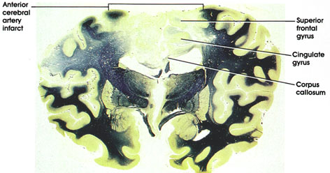
This is a coronal section of the brain showing bilateral anterior cerebral artery infarcts. The infarcts involve structures supplied by the anterior cerebral artery such as the superior frontal gyrus, the cingulate gyrus, and the corpus callosum. Patients with this type of infarct will show bilateral lower extremity weakness or paralysis associated with upper motor neuron signs in the affected extremities. The upper extremities and the face will be spared in this type of lesion because their cortical areas are supplied by the middle cerebral artery.
ARTERY INFARCT
Coronal
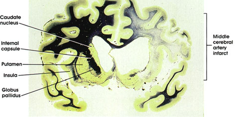
This is a coronal section of the brain at the level of the corpus striatum, showing a middle cerebral artery infarct. The structures involved in the infarct are those supplied by the middle cerebral artery and include, at this level, the caudate nucleus, internal capsule, putamen, insula, globus pallidus, and the lateral surface of the hemisphere. Patients with this type of infarct will show contralateral weakness or paralysis of the upper motor neuron variety involving mainly the face and upper extremities. The lower extremity will be largely spared because its area of cortical representation is supplied by the anterior cerebral artery. Middle cerebral artery infarcts in the dominant left hemisphere will also be associated with disturbance in language known as aphasia.
Axial
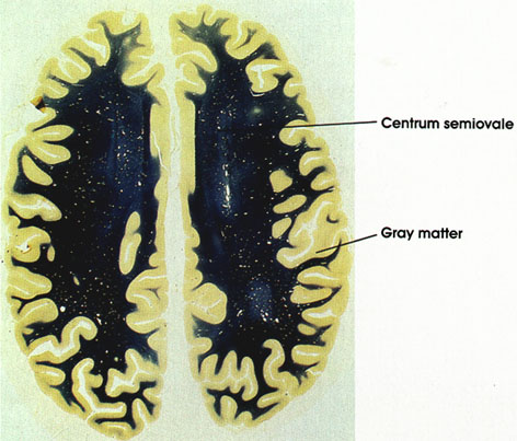
Centrum semiovale: White matter core of the cerebral hemispheres. Contains myelinated nerve fibers entering or leaving the cerebral hemispheres.
Gray matter: Contains neurons whose axons contribute to the centrum semiovale.
Axial
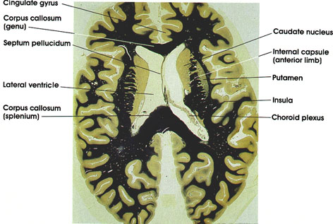
Cingulate gyrus: Located dorsal to the corpus callosum. A component of the limbic lobe, which also includes the subcallosal gyrus, isthmus, parahippocampal gyrus and uncus.
Caudate nucleus: C-shaped mass of gray matter closely related to the lateral ventricle. A component of the basal ganglia and thus plays a role in motor control.
Internal capsule (anterior limb): Separates the caudate nucleus and cerebral cortex.
Putamen: One of the basal ganglia nuclei. Located lateral to the anterior limb of the internal capsule. Concerned with motor control.
Insula: Also referred to as the island of Reil, lies deep in the sylvian fissure. Concerned with autonomic function.
Choroid plexus: Vascular pial fold in the body of the lateral ventricle. One of the sites of formation of cerebrospinal fluid.
Corpus callosum: C-shaped bundle of heavily myelinated fibers connecting the two cerebral hemispheres. Important in interhemispheric transfer of information. Section is through the genu (anterior) and splenium (posterior) parts of the corpus callosum.
Lateral ventricle: Note the characteristic bulge of the caudate into the cavity of the lateral ventricle.
Septum pellucidum: Separates the two lateral ventricles. May contain a cavity, cavurn septi pellucidi
Axial
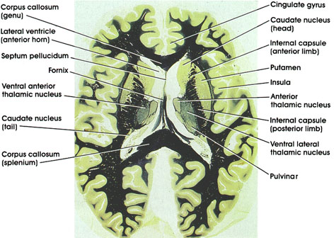
Corpus callosum: C-shaped bundle of heavily myelinated fibers. Connects the two hemispheres. Important in interhemispheric transfer of information. Section is through the genu (anterior) and splenium (posterior) parts of the corpus callosum.
Cingulate gyrus: A component of the limbic lobe. Located dorsal to the corpus callosurn on the medial surface of the hemisphere.
Caudate nucleus: C-shaped mass of gray matter closely related to the lateral ventricle. A component of the basal ganglia and thus concerned with motor control. Section shows the head of the caudate (anterior) and the much smaller tail (posterior).
Internal capsule: A massive bundle of myelinated fibers conveying motor and sensory impulses from and to the cerebral cortex. The anterior limb separates the caudate nucleus and putamen, whereas the posterior limb separates the putamen and thalamus.
Putamen: A component of the basal ganglia. Concerned with motor control.
Insula: Also referred to as the island of Reil. Lies deep in the sylvian fissure. Concerned with autonomic function.
Anterior thalamic nucleus: Belongs to the anterior group of thalamic nuclei. Has reciprocal connections with the mamillary body via the mamillothalamic tract and with the cingulate gyrus via the internal capsule. Considered part of the limbic system and thus plays a role in emotional behavior and memory.
Ventral lateral thalamic nucleus: Traversed by bundles of myelinated fibers. Belongs to the lateral group of thalamic nuclei. Relay thalamic nucleus for cerebellar fibers to the motor cortex. Reciprocally connected with the primary motor cortex. Concerned with motor control.
Pulvinar: One of the lateral group of thalamic nuclei. Has reciprocal connections with the medial and lateral geniculate bodies caudally and the association parietal, temporal, and occipital cortices rostrally. involved in several neural functions, including vision, audition, speech, and pain.
Fornix: A paired C-shaped fiber tract connecting the hippocampus with several brain regions, including the mamillary body, anterior thalamic nucleus, septal nuclei, and cingulate gyrus.
Lateral ventricle (anterior horn): Note characteristic bulge of head of caudate into the anterior horn.
Septum pellucidum: Separates the two cavities of the lateral ventricle. May contain a cavity, cavurn septi pellucidi.
Ventral anterior thalamic nucleus: One of the lateral group of thalamic nuclei. Characteristically traversed by heavily myelinated fiber bundles. Receives fibers from the basal ganglia and projects to the motor cortex. The posterior limb separates the thalamus from the putamen
Axial
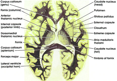
Corpus callosum: C-shaped bundle of heavily myelinated fibers. Connects the two hemispheres. Important in interhemispheric transfer of information. Section shows the genu (anterior) and splenium (posterior) parts of the corpus callosum.
Caudate nucleus: C-shaped mass of gray matter closely related to the lateral ventricle. A component of the basal ganglia and thus concerned with motor control. Section shows the head of the caudate (anterior) and the much smaller tail (posterior).
Putamen: A component of the basal ganglia. Plays a role in motor control. Medial to the external capsule and lateral to the caudate and globus pallidus.
Globus pallidus: One of the basal ganglia nuclei. Located medial to the putamen. The posterior limb of the internal capsule separates it from the thalamus. Traversed by heavily myelinated fiber bundles. Receives fibers from the caudate and putamen and projects to the thalamus (ventral anterior nucleus). Has reciprocal connections with the subthalamic nucleus. Plays a role in motor control.
External capsule: An efferent cortical fiber bundle. Lateral to the putamen.
Claustrum: A thin zone of gray substance located between the external and extreme capsules.
Extreme capsule: An efferent cortical fiber bundle. Situated lateral to the claustrum.
Stria medullaris thalami: A bundle of myelinated fibers connecting the septal nuclei with the habenular complex. Characteristically located dorsomedial to the thalamus.
Fimbria of fornix: Efferent fibers from the hippocampus. Continues as the body of the fornix.
Fornix: C-shaped, paired fiber system connecting the hippocampus with several brain regions, including the mamillary body, anterior thalamic nucleus, septal nuclei, and cingulate gyrus. The part of the fornix seen here represents the columns of the fornix.
Anterior thalamic nucleus: Belongs to the anterior group of thalamic nuclei. Has reciprocal connections with the mamillary body via the mamillothalamic tract and with the cingulate gyrus via the internal capsule. Considered part of the limbic system. Plays a role in emotional behavior and memory.
Internal capsule (posterior limb): Separates the putamen from the thalamus. Massive fiber bundle conveying impulses to and from the cerebral cortex. Lesions in internal capsule result in contralateral motor and sensory deficits.
Dorsomedial thalamic nucleus: Belongs to the medial group of thalamic nuclei. Most highly developed in man. Located medial to the internal medullary lamina. Has reciprocal connections with the prefrontal cortex and hypothalamus. Receives input from other thalamic nuclei. Concerned with affective behavior and memory.
Forceps major: Extension of the splenium of the corpus callosurn posteriorly into the occipital lobes.
Lateral ventricle: Extension of the cavity of the lateral ventricle into the occipital lobe (occipital horn).
Axial
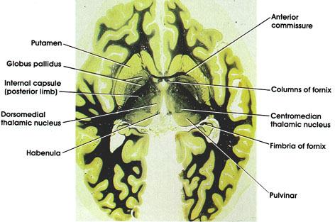
Columns of fornix: Body of fornix separates into two columns, each of which curves ventrally and courses through the hypothalamus on its way to the mamillary body.
Anterior commissure: A compact fiber bundle in close proximity to anterior extent of the fornix. Interconnects olfactory bulbs and the temporal cortices.
Centromedion thalamic nucleus: Belongs to the intralaminar group of thalamic nuclei. Receives fibers from several sources, motor and sensory, that project diffusely to cerebral cortex either directly or indirectly via other thalamic nuclei. Plays a role in cortical arousal response and in pain mechanism.
Pulvinar: Belongs to the lateral group of thalamic nuclei. Has reciprocal connections with the medial and lateral geniculate bodies caudally and the association parietal, temporal, and occipital cortices rostrally. Plays a role in several neural functions, including vision, audition, speech, and pain.
Fimbria of fornix: Efferent fibers from the hippocampus that merge with the fornix.
Putamen: Lateral to globus pallidus. One of the basal ganglia nuclei. Concerned with motor control.
Globus pallidus: Medial to the putamen. Separated from the thalamus by the posterior limb of the internal capsule. Has two segments: outer, close to the putamen, and inner, close to the internal capsule. Characterized by heavily myelinated fibers traversing it. A component of the basal ganglia. Concerned with motor control.
Internal capsule (posterior limb): Separates putamen and globus pallidus from thalamus. Carries motor and sensory fibers from and to the cerebral cortex.
Dorsomedial thalamic nucleus: Belongs to the medial group of thalamic nuclei. Most highly developed in man. Concerned with affective behavior and memory.
Habenula: A nuclear mass located dorsal to the thalamus at the junction of the diencephalon and midbrain. A component of the limbic system.
Axial
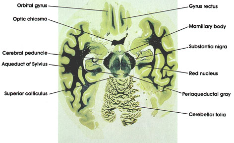
Gyrus rectus: inferior surface of the frontal lobe. Medial to orbital gyrus.
Mamillary body: A pair of spherical nuclear masses caudal to the optic chiasma and protruding from the ventral surface of the posterior hypothalamus in the interpeduncular fossa (between the cerebral peduncles). Concerned with memory function.
Substantia nigra: A pigmented (melanin) nuclear mass located in the base of the midbrain above the cerebral peduncles. invariably the site of pathologic changes in Parkinson's disease.
Red nucleus: So-called because of a pinkish color in the fresh state owing to its high vascularity. Links the cerebellum, cerebral cortex, and spinal cord.
Periaqueductal gray: A nuclear mass surrounding the aqueduct of Sylvius. Related to pain mechanisms.
Cerebellar folia: Leaf-like folds of the cerebellar cortex separated from each other by sulci and supported by a core of white matter.
Aqueduct of Sylvius: Connects the fourth with the third ventricle.
Cerebral peduncle: Descending corticofugal fiber system. Lesion results in contralateral muscle weakness or paralysis.
Superior colliculus: Cellular mass concerned with visual reflexes.
Axial
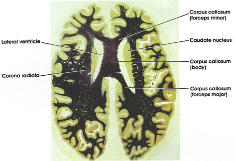
Corpus callosum: C-shaped massive bundle of heavily myelinated fibers. Connects the two hemispheres. Important in interhemispheric transfer of information. Figure shows the body of the corpus callosurn and extensions into the frontal pole (forceps minor) and into the occipital pole (forceps major).
Caudate nucleus: Another C-shaped mass of gray matter closely related to the lateral ventricle. A component of the basal ganglia. Important for motor control.
Lateral ventricle: Contains cerebrospinal fluid. Note characteristic bulge of caudate nucleus into the cavity of the lateral ventricle.
Corona radiata: White matter core of the cerebral hemispheres. Contains afferent and efferent fibers to and from the cerebral cortex.
Parasagittal
Centrum semiovale: White matter core of the cerebral hemispheres. Contains myelinated nerve fibers entering or leaving the cerebral hemispheres.
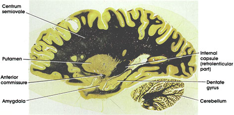
Putamen: One of the basal ganglia nuclei. Concerned with motor control. Note that at this most lateral parasagittal section, only the putamen of the basal ganglia nuclei is seen. In subsequent more medial sections, both the caudate nucleus and globus pallidus will also be visible.
Anterior commissure: A compact fiber bundle ventral to putamen. Interconnects olfactory bulbs and the temporal cortices.
Amygdala: From the Greek word amygdala, almond. The nuclei resemble almonds in shape and are located in the tip of the temporal lobe beneath the cortex of the uncus. Part of the limbic system and intimately connected with different components of the limbic system. Concerned with a variety of functions related to the limbic system such as emotional behavior, food intake, arousal, sexual activity, and a variety of motor activities.
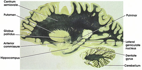
Cerebellum: Located underneath the posterior part of the cerebral hemisphere.
Dentate gyrus: So-named because of its toothed or beaded surface appearance. A component of the hippocampal formation. Occupies the interval between the hippocampus and the parahippocampal gyrus.
Internal capsule: A component of the internal capsule, known as the retrolenticular part and characteristically located behind the putamen, is seen here. Note the different components of the internal capsule (anterior limb, posterior limb) in other sections. The retrolenticular component of the internal capsule contains the visual radiation, corticotectal, corticonigral, and corticotegmental fibers.
Parasagittal
Centrum semiovale: White matter core of the cerebral hemispheres. Contains myelinated nerve fibers entering or leaving the cerebral hemispheres.
Putamen: One of the basal ganglia nuclei. Concerned with motor control. Note that this section also shows the globus pallidus, another component of the basal ganglia.
Globus pallidus: Another of the basal ganglia nuclei. Notice the difference in appearance from putamen. Globus pallidus is traversed by heavily myelinated fiber bundles. Note relationship to the anterior commissure.
Anterior commissure: A compact fiber bundle in close proximity to globus pallidus. Interconnects the olfactory bulbs and the temporal cortices.
Hippocampus: From the Greek word hippocampos, seahorse. The hippocampus is a major component of the limbic system. In man, the hippocampus is the largest component of the hippocampal formation. It plays an important role in memory function.
Cerebellum: Located ventral to the posterior part of the cerebral hemisphere.
Dentate gyrus: Another component of the hippocampal formation. So-named because of its toothed or beaded surface appearance. Occupies the interval between the hippocampus and the parahippocampal gyrus.
Lateral geniculate nucleus: One of the thalamic nuclei. Concerned with visual function. Receives fibers from the optic tract and projects into the primary visual cortex via the geniculocalcarine fiber pathway.
Pulvinar: One of the lateral group of thalamic nuclei. Has reciprocal connections with the medial and lateral geniculate bodies caudally and the association parietal, temporal, and occipital cortices rostrally. Involved in several neural functions, including vision, audition, speech, and pain.
AND LATERAL GENICULATE NUCLEUS
Parasagittal
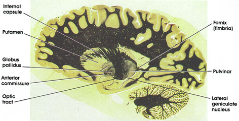
Internal capsule: A major fiber pathway carrying fibers from and to the cerebral cortex.
Putamen: One of the basal ganglia nuclei, concerned with motor control. Note relationship to globus pallidus.
Globus pallidus: Another of the basal ganglia nuclei. The two components of globus pallidus are shown in this section, an outer and an inner component. Note the difference in appearance of globus pallidus and putamen. Globus pallidus is characteristically traversed by heavily myelinated fiber bundles.
Anterior commissure: Compact fiber bundle inclose proximity to globus pallidus. interconnects the olfactory bulbs and the temporal cortices.
Optic tract: A bundle of heavily myelinated fibers conveying impulses from the retinae to the lateral geniculate nucleus as well as to the pretectal area.
Lateral geniculate nucleus: One of the thalamic nuclei. Concerned with visual functions. Receives fibers from the optic tract and projects to the primary visual cortex via the geniculocalcarine fiber pathway.
Pulvinar: One of the lateral group of thalamic nuclei. Has reciprocal connections with the medial and lateral geniculate bodies caudally and the association parietal, temporal, and occipital cortices rostrally. Involved in several neural functions, including vision, audition, speech, and pain.
Fornix (fimbria): Outflow tract from the hippocampus. When traced posteriorly on the floor of the inferior horn of the lateral ventricle, the fimbria continues as the crus of the fornix, which begins beneath the splenium of the corpus callosurn (see Plates 362, 363 and 364). The fornix connects the hippocampus with several brain regions, including the mamillary body, anterior thalamic nucleus, septal nuclei, and cingulate gyrus.
AND MEDIAL GENICULATE NUCLEUS
Parasagittal
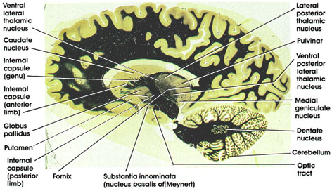
Ventral lateral (thalamic) nucleus: One of the lateral group of thalamic nuclei. Note the heavy bundle of myelinated fibers traversing the nucleus. Receives fibers from the cerebellum via the brachium conjunctivum. Has reciprocal connections with the primary motor cortex. Plays a role in motor control.
Caudate nucleus: One of the basal ganglia nuclei. Note its characteristic bulge into the cavity of the lateral ventricle. Concerned with motor control. This section shows in addition the putamen and globus pallidus.
Internal capsule (genu): The part of the internal capsule between the anterior and posterior limbs. Contains corticobulbar fibers, which terminate upon motor nuclei of the brain stem.
Internal capsule (anterior limb): Separates the caudate nucleus and putamen.
Globus pollidus: One of the basal ganglia nuclei. Characterized by heavily myelinated bundles of nerve fibers coursing through it. Note the difference in appearance between globus pallidus and both the caudate and putamen. Both components of the globus pallidus are shown in this section, the lateral (outer) and the medial (inner) segments. Concerned with motor control.
Putamen: Another of the basal ganglia nuclei. Similar in structure to caudate nucleus. Together with caudate and globus pallidus, it constitutes the corpus striatum.
Internal capsule (posterior limb): This part of the internal capsule separates the thalamus from the basal ganglia. The posterior limb of the internal capsule contains corticospinal, corticorubral, corticothalamic, and thalamocortical fibers.
Substantia innominata (nucleus basalis of Meynert): Located ventral to internal capsule and anterior commissure. The substantia innominata contains the nucleus basalis of Meynert, the neurons of which are rich in acetylcholine. Neurons in this nucleus project diffusely to the cerebral cortex and are believed to be involved in Alzheimer's disease. Alzheimer was a nineteenth-century German neurologist.
Optic tract: Carrying fibers from the retinae to the lateral geniculate nucleus and the pretectal area.
Cerebellum: Located ventral to the posterior part of the cerebral hemisphere.
Dentate nucleus: The largest of the deep cerebellar nuclei. Receives fibers from the Purkinje neurons in the hemispheres of the cerebellum and projects to the thalamus via the dentatorubrothalamic fiber system. Purkinje was a nineteenth-century Bohemian anatomist and physiologist.
Medial geniculate nucleus: One of the thalamic nuclei. Concerned with audition. Receives auditory fibers from the brain stem and projects to the primary auditory cortex in the temporal lobe.
Ventral posterior lateral (thalamic) nucleus: One of the lateral group of thalamic nuclei. Receives fibers of the medial lemniscus and spinothalamic tract. Reciprocally connected with the primary somesthetic cortex.
Fornix (fimbria): Axons of hippocampal neurons, continuous with the crus of the fornix. The fornix connects the hippocampus with several brain regions, including the mamillary body, anterior thalamic nucleus, septal nuclei, and cingulate gyrus.
Pulvinar: One of the lateral group of thalamic nuclei. Reciprocally connected with the medial and lateral geniculate bodies caudally and the association parietal, temporal, and occipital cortices rostrally. Involved in several neural functions, including vision, audition, speech, and pain.
Lateral posterior (thalamic) nucleus: One of the lateral group of thalamic nuclei. The borderline between this nucleus and the pulvinar is vague; the term pulvinar-lateral posterior complex has been used to refer to this nuclear complex.
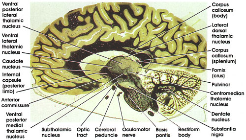
Parasagittal
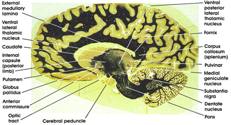
Ventral lateral (thalamic) nucleus: One of the lateral group of thalmic nuclei. Characteristically traversed by heavily myelinated fiber bundles. Receives fibers from the cerebellum and is reciprocally connected with the primary motor cortex. Concerned with motor control.
Internal capsule (posterior limb): Contains fibers destined for and leaving the cerebral cortex. This part of the internal capsule separates the thalamus from the basal ganglia.
Caudate nucleus: One of the basal ganglia nuclei. Concerned with motor function. Note the similarity in appearance between caudate and putamen.
Globus pallidus: Another of the basal ganglia nuclei. Characterized by heavily myelinated bundles traversing it. The two components of globus pallidus, the outer and inner segments, are seen in this section. Note proximity to anterior commissure.
Putamen: One of the basal ganglia nuclei. Concerned with motor function.
Anterior commissure: Compact fiber bundle located in close proximity to globus pallidus. interconnects the olfactory bulbs and the temporal cortices.
Optic tract: Myelinated fiber bundle conveying visual impulses from the contralateral field of vision to the lateral geniculate nucleus and the pretectal area.
Pons: Note the characteristic ventral bulge known as the basis pontis.
Cerebral peduncle: Located ventral to the substantia nigra. Contains descending corticofugal fibers, including the corticospinal, corticobulbar, and corticopontine fiber systems. Lesions result in weakness (paresis) or paralysis of the contralateral half of the body, including the face.
Dentate nucleus: One of the deep cerebellar nuclei. Receives fibers from Purkinje neurons in the hemispheres of the cerebellum and projects to the thalamus (ventral lateral nucleus) and red nucleus via the dentatorubro thalamic system.
Substantia nigra: A large nuclear mass in the midbrain. Contains pigmented cells (melanin). Connected with the basal ganglia and thalamus. Important in motor control. This area is invariably the site of pathological changes associated with Parkinson's disease.
Medial geniculate nucleus: One of the thalamic nuclei. Concerned with audition. Receives auditory fibers from the brain stem and is reciprocally connected with the primary auditory cortex.
Pulvinar: One of the lateral group of thalamic nuclei. Reciprocally connected with the medial and lateral geniculate bodies caudally and the association parietal, temporal, and occipital cortices rostrally. Involved in several neural functions, including vision, audition, speech, and pain.
Corpus callosum (splenium): The caudal part of the corpus callosum. Important in transfer of visual information between the two hemispheres.
Fornix (crus): Continuation of the same system seen in other sections. The fimbria of the fornix arising from the hippocampus continues as the crus. Note relationship to splenium of corpus callosum.
Ventral posterior lateral (thalamic) nucleus: One of the lateral group of thalamic nuclei. Receives the medial lemniscus and the spinothalamic tract and projects to the primary somesthetic cortex in the postcentral gyrus.
External medullary lamina: A bundle of myelinated fibers separating the medial from the lateral group of thalamic nuclei.
Parasagittal
Internal capsule (posterior limb): The part of the internal capsule separating the thalamus from the basal ganglia. Contains fibers destined for and leaving the cerebral cortex.
Caudatenucleus: One of the basal ganglia nuclei. Concerned with motor control. Note the characteristic bulge into the cavity of the lateral ventricle.
Anterior commissure: A compact fiber bundle connecting the olfactory bulbs and the temporal cortices.
Ventral posterior medial thalamic nucleus: One of the lateral group of thalamic nuclei. Receives the trigeminothalamic fiber system, including taste fibers, and projects to the primary somesthetic cortex in the postcentral gyrus.
Optic tract: Conveys impulses from the contralateral visual field to the lateral geniculate nucleus and the pretectal area.
Cerebral peduncle: A bundle of corticofugal fibers located inferior to the substantia nigra in the midbrain. Contains corticospinal, corticobulbar, and corticopontine fiber bundles. Lesions produce weakness or paralysis in the contralateral half of the body, including the face.
Oculomotor nerve: Seen leaving the ventral surface of the midbrain.
Subthalamic nucleus: Also known as corpus Luysii. Shaped like a biconcave lens. Receives fibers from and projects to the globus pallidus. Discrete lesions here result in an abnormal type of flinging movement known as ballism.
Basis pontis: Contains descending corticofugal fibers, pontine nuclei, and pontocerebellar fibers.
Restiform body: Also known as inferior cerebellar peduncle. A compact bundle of nerve fibers connecting the medulla with the cerebellum. Tracts and fibers forming this bundle originate in the medulla and the spinal cord.
Dentate nucleus: One of the deep cerebellar nuclei. Receives fibers from the Purkinje neurons of the cerebellar hemispheres and projects via the dentatorubrothalarnic system to the contralateral red nucleus and ventrolateral nucleus of the thalamus. Concerned with motor control.
Substantia nigra: The largest nuclear mass in the midbrain. Contains pigmented neurons (melanin). Connected with basal ganglia and thalamus. important in motor control. This area is invariably the site of pathological changes associated with Parkinson's disease.
Centromedian (thalamic) nucleus: Belongs to the intralaminar group of thalamic nuclei. Concerned with a variety of sensory and motor functions and arousal.
Pulvinar: One of the lateral group of thalamic nuclei. Has reciprocal connections with the medial and lateral geniculate bodies caudally and association parietal, temporal, and occipital cortices rostrally. involved in several neural functions, including vision, audition, speech, and pain.
Corpus callosum (splenium): The posterior part of the corpus callosurn. Concerned with transfer of visual information between the hemispheres.
Fornix (crus): Note the relationship of the crus of the fornix to the splenium of the corpus callosurn. The crus is a continuation of the fimbria of the fornix (see Plates 354, 355, 360, and 361). Rostrally, the crus continues as the body of the fornix. The fornix connects the hippocampus with several brain regions, including the mamillary body, anterior thalamic nucleus, septal nuclei, and cingulate gyrus.
Corpus callosum (body): The largest part of the corpus callosum. Between the splenium and the genu. Important in interhemispheric transfer of information.
Lateral dorsal (thalamic) nucleus: One of the dorsal subgroup of the lateral group of thalamic nuclei. An association thalamic nucleus.
Ventral posterior lateral (thalamic) nucleus: One of the lateral group of thalamic nuclei. Receives the medial lemniscus and the spinothalamic tract and is reciprocally connected with the somesthetic cortex of the postcentral gyrus.
Ventral lateral (thalamic) nucleus: Another of the lateral group of thalamic nuclei. Concerned with motor function. Characteristically traversed by heavily myelinated fiber bundles. Receives fibers from deep cerebellar nuclei and is reciprocally connected with the primary motor cortex.
Midsagittal
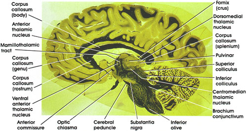
Mamillothalamic tract: A myelinated fiber bundle connecting the mamillary body with the anterior thalamic nucleus. Seen here approaching the anterior thalamic nucleus.
Corpus callosum (genu): The part of the corpus callosurn between the rostrum and the body. important in interhemispheric transfer of information.
Ventral anterior thalamic nucleus: One of the lateral group of thalamic nuclei. Concerned with motor function. Characteristically traversed by heavily myelinated fiber bundles. Receives fibers from the basal ganglia and projects to the motor cortex.
Corpus callosum (rostrum): The most rostral part of the corpus callosum. Concerned with interhemispheric transfer of information.
Anterior commissure: A compact bundle of myelinated fibers. Interconnects olfactory bulbs and the temporal cortices.
Optic chiasma: Site of partial crossing of optic nerve fibers before formation of the optic tracts. Lesions in the optic chiasma result in a characteristic visual field loss known as bitemporal hemianopia.
Cerebral peduncle: A bundle of corticofugal fibers located ventral to the substantia nigra in the midbrain. Contains corticospinal, corticobulbar, and corticopontine fibers. Lesions result in contralateral weakness or paralysis, including the face.
Substantia nigra: A large nuclear mass in the ventral part of the-midbrain. Located dorsal to the cerebral peduncles. Melanin-containing neurons in this nucleus are connected with the basal ganglia and the thalamus. The substantia nigra is an important structure in the genesis of movement. Invariably the site of pathology in Parkinson's disease.
Inferior Olive: Located in ventral part of the medulla oblongata. Receives cortical and subcortical fibers and projects to the cerebellum via the restiform body.
Brachium conjunctivum: Also known as superior cerebellar peduncle. Contains axons of deep cerebellar nuclei destined for the red nucleus and thalamus.
Inferior colliculus: An elevation on the dorsal surface of the midbrain. Contains neurons related to the auditory system.
Superior colliculus: An elevation in the dorsal surface of the midbrain. Related to the visual system.
Centromedian (thalamic) nucleus: Belongs to the intralaminar group of thalamic nuclei. Related to a variety of sensory and motor functions as well as arousal.
Corpus callosum (splenium): The most caudal part of the corpus callosum. Concerned with transfer of visual information between the hemispheres.
Pulvinar: One of the lateral group of thalamic nuclei. Reciprocally connected with the medial and lateral geniculate nuclei caudally and the association parietal, temporal, and occipital cortices rostrally. Plays a role in several neural functions, including vision, audition, speech, and pain.
Fornix (crus): Continuation of the fimbria of the fornix. Continues rostrally as the body of the fornix. The fornix connects the hippocampus with several brain regions, including the mamillary body, anterior thalamic nucleus, septal nuclei, and cingulate gyrus.
Dorsomedial (thalamic) nucleus: One of the medial group of thalamic nuclei. Most highly developed in man. Has reciprocal connections with the prefrontal cortex and hypothalamus. Receives input from other thalamic nuclei. Concerned with affective behavior and memory.
Anterior (thalamic) nucleus: Belongs to the anterior group of thalamic nuclei. Has reciprocal connections with the mamillary body via the mamillothalarnic tract and with the cingulate gyrus via the internal capsule. Considered part of the limbic system and plays a role in emotional behavior and memory.
Corpus callosum (body): The largest part of the corpus callosum. Between the genu and the splenium. Concerned with interhemispheric transfer of information.