Section 15: Endocrine Glands
These cellular masses, designated as the ductless glands or glands of internal secretion, have during development lost their original connection with the epithelium of the free surface. Their secretions are called hormones. The gland cells produce specific chemical substances, which are secreted in a rich capillary bed and carried by the blood to another part, or parts, of the body where they have a distinctive function. The endocrine glands are essentially a vertebrate development, and any one of these hormonal substances has a similar action in all vertebrates with little or no species-specific function.
Endocrine glands may appear as distinct organs (e.g., the hypophysis and adrenal glands), may be found associated with exocrine glands (e.g., pancreatic islets and the interstitial cells of the testes), may appear as mixed endocrine glands (e.g., the thyroid and parathyroid glands), or may have cells so diffusely distributed that they are not usually considered as organs (e.g., argentaffin cells of the digestive system).
Some endocrine glands are essential for life; these include the adrenal cortex, pancreatic islets, and the parathyroid glands. The other endocrine glands, although not essential for life, determine to a great extent the quality of ones life and the ability to adapt to stress. The endocrine glands, separately and in conjunction with the nervous system, are coordinators of body functions that maintain the organism in a viable homeostatic state.
The structural/functional organization of the endocrine glands is diverse but distinctive. In general, all endocrine glands store their secretory products either within the cells of origin or within cellular follicles or sacs. The cells of the adrenal cortex contain minimal amounts of stored hormone, whereas in the pancreas and pituitary gland (hypophysis), secretory granules (if preserved) are usually evident. In the thyroid gland, the hormone is stored extracellularly in a pool surrounded by epithelial gland cells (a follicle). In this case, the release of the hormone into the blood stream involves the reabsorption and transfer of the hormone through the cells of origin into the extracellular space, where it enters the capillaries.
An essential feature of the endocrine glands is the manner in which the secretory activity is regulated by a feedback mechanism. As an example, the beta cell of the anterior lobe of the hypophysis secretes adrenocorticotropic hormone (ACTH), which stimulates the secretion of certain hormones from the adrenal cortex. As the level of adrenal cortical hormones rises in the blood stream, the secretion of ACTH is inhibited. Declining levels of the hormones of the adrenal cortex result in an increased secretion of ACTH by the pituitary. In this manner, appropriate levels of adrenal cortical hormones are maintained in the blood stream.
Specific details of the structure and function of the endocrine glands will be found in this section and, as appropriate, in the sections concerned with the digestive, urinary, male reproductive, female reproductive, and nervous systems.
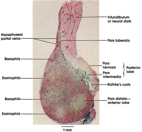
Several features of the hypophysis can be seen in this low-power photomicrograph. Elements of the infundibulum or neural stalk, which develops from the floor of the diencephalon, and the derivatives of Rathke's* pouch, which arise from an outpocketing of ectoderm from the roof of the mouth of the embryo, are shown. The pouch derivatives include the pars distalis and pars intermedia. Note the cyst- like clefts between the pars distalis and the pars intermedia, which are developmental remnants of the primitive cavity of Rathke's pouch. Elements of the important hypophysealportal blood circuit can be seen in the pars tuberalis.
Even at this low magnification, clusters of basophils and eosinophils can be seen in the pars distalis, permitting their study at higher magnifications.
Pituitary Gland
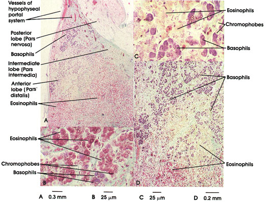
A. 32 x, B. 316 x, C. 316 x D. 82 x.
This plate is at higher magnification than the preceding one. This permits easier location and identification of various cell types that populate the pituitary gland. One may find three cell types grouped under two headings: (1) chromophils, which include eosinophils (two types in this preparation) and basophils, and (2) chromophobes.
it must be remembered that empirical histological methods cannot differentiate precisely cell types. This can now be done by immunocytochernistry, but this method effaces other structural detail; therefore, several methods are usually required. Students interested in the complexities of cell typing should consult recent original investigative references. Nevertheless, one can locate populations of cells whose functions can be ascribed because they bear consistent staining characteristics.
Eosinophils are of two types and are known to secrete growth hormone (somatotropin) and prolactin. Both cells are stainable with orange G, but not with the periodic acid-Schiff method.
Basophils fall into three types: one type produces follicle-stimulating hormone (FSH) and luteinizing hormone (LH); a second type produces thyrotropic hormone; and a third type produces adrenocorticotropic (ACTH) hormone. These cells do not stain with orange G but do stain with the periodic acid-Schiff method, reflecting the glycoprotein, rather than any acid, nature of the synthesized secretory product.
Some chromophobes may be supporting cells with long branching processes forming a network within the parenchyma of the organ. Other chromophobe cells are not numerous and may be either transitionally degranulated cells or may be considered as reserve cells.
The pituitary gland receives its blood supply from the internal carotid artery and the superior and inferior hypophyseal arteries. The superior artery becomes a primary capillary plexus with fenestrated capillaries. Neurosecretory neurons contain releasing or inhibitory factors and are intimately associated with the capillary plexuses. These capillary plexuses rejoin to form portal (vessels located between and joining two capillary beds) veins, which once again form capillary plexuses around cells of the adenohypophysis (pars distalis). It can be appreciated that this system is critically important to normal hypophyseal function.
Pituicyte nuclei: The pituicyte perikaryon is not seen. These cells constitute 25 to 30 percent of the posterior lobe of the hypophysis. Function is poorly understood. Pituicytes are related to the neurosecretory axons and they correspond to neuroglia of the brain.
Capillary: Capillaries receive hormones secreted from Herring* bodies for distribution throughout the body. The posterior lobe of the hypophysis is highly vascularized.
Anterior, intermediate, and posterior lobes
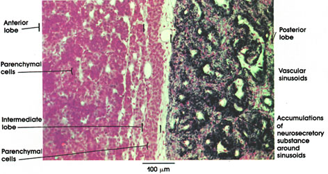
Gomori's chrome alum hematoxylin and phloxine stains, 162 x.
The anterior lobe is made of anastomosing cords and plates separated by capillaries. Several trophic hormones are produced in this lobe: somatotropin, follicle-stimulating hormone, luteinizing hormone, luteotropic hormone (prolactin), thyrotropic hormone, and adrenocorticotropic hormone.
The intermediate lobe is sandwiched between the anterior and posterior lobes. It has colloid-filled cysts. it elaborates melanocyte-stimulating hormone in some species (amphibia). In man and other mammals, the hormone appears to influence melanin synthesis.
The posterior lobe is made of sheets of cells and is rich in neurosecretory material. The latter is concentrated around the sinusoids. Two hormones have been extracted from this neurosecretory material, antidiuretic hormone and oxytocin.
Anterior lobe
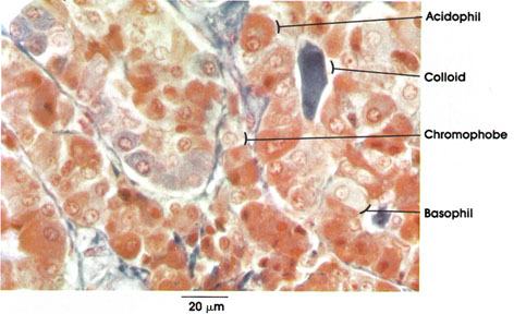
Chromophobe: Small, poorly staining cell. The cytoplasm is scanty and devoid of granules. Cell contours are rounded or polygonal. Also known as reserve or chief cell.
Acidophil: Larger than a chromophobe. Cytoplasm rich in granules, which stain with acid dyes. Since they also take basic dyes, a better name might be alpha cell. Secrete growth and lactogenic hormones. The lactogenic hormone- producing acidophils increase during pregnancy. Excessive use or production of growth hormone as occurs in pituitary tumors produces gigantism if it occurs before puberty and acromegaly if it occurs after puberty.
Basophil: Beta cell. Larger than the average acidophil and less heavily granulated. Secretes thyroid- stimulating hormone (TSH), adrenocorticotropic hormone (ACTH), follicle-stimulating hormone (FSH), and luteinizing hormone (LH).
Colloid: Secretion product. Found in center of some cords.
Anterior lobe
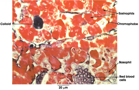
Colloid: Secretion product. Found in the center of some cords.
Chromophobe: Small, faintly staining and less numerous than other cells in the pituitary. Scanty cytoplasm devoid of granules. Tend to cluster near center of cords.
Eosinophils: Alpha cells. Larger than chromophobes. Cytoplasm filled with acidophilic granules. Source of growth and lactogenic hormones.
Basophil: Beta cell. Larger than the average eosinophil and less heavily granulated. Secretes thyroid- stimulating hormone, adrenocorticotropic hormone, follicle-stimulating hormone, and luteinizing hormone.
Red blood cells: Filling the sinusoids.
Posterior lobe Herring body
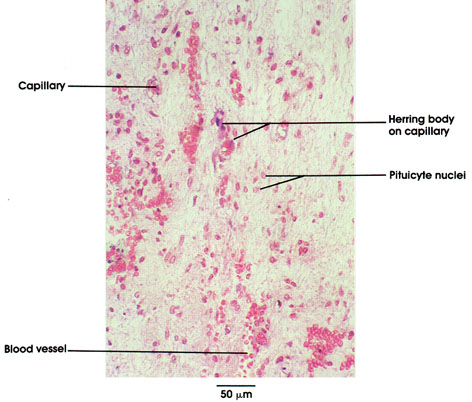
Herring* body: Herring bodies represent dilated terminal portions of neurosecretory axons constituting the hypothalamohypophyseal tract. They are always in close proximity to capillaries. Within the dilatations of various sizes are neurosecretory granules, which stain blue in this preparation. The Herring body granules consist of hormone precursors for either oxytocin or vasopressin, plus a binding protein for each hormone and adenosine triphosphate (ATP). The neurosecretory material is synthesized in neurons located in the hypothalamus and transported down the axons of the hypothalamohypophyseal tract to reach the axon terminals (Herring bodies). Most of the vasopressin is thought to be synthesized in neurons located in the supraoptic nucleus and oxytocin in neurons of the paraventricular nucleus. Vasopressin (pitressin, 13-hypophamine) is an antidiuretic hormone and in pharmacologic doses causes smooth muscle to contract, particularly those in blood vessels. Oxytocin (pitocin, A-hypophamine) causes myometrial contractions at term and promotes milk release during lactation.
*Herring bodies also contain synaptic vesicles 40 to 60 nm in diameter that are similar in structure to those found in cholinergic synapses. Their function here is not known.
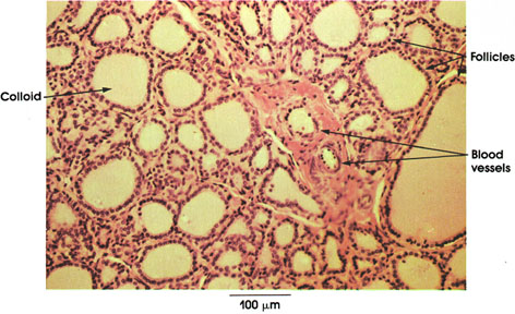
Follicles: Structural units of the thyroid gland. Note variations in shape (rounded or tubular) and size (0.05 to 0.5 mm in diameter). Close packing with a thin reticular network between adjacent follicles. Single layer of cells forms a hollow sphere. Nucleus centrally or basally placed.
Colloid: Found in the lumen of follicles. Chemical composition is a glycoprotein-iodine complex (thyroglobulin). Of the several iodinated compounds found in the gland the 3,5,3-triiodothyronine is hormonally the most active. The follicles release about 100 mg of hormone daily. Normal thyroid function is essential for the normal growth, development, and well-being of man and animals. Hypofunction of the thyroid in infants results in cretinism, characterized by dwarfism, mental deficiency, slow heart rate, muscular weakness, and gastrointestinal disturbances. Thyroid hormone given to infants at an early stage of cretinism can alleviate the symptoms. In adults, hypothyroidism results in muscular weakness, mental slowing, cold intolerance, and reflex and skin changes. When the hormone is produced in excess (hyperthyroidism), excessive appetite and thirst, weight loss, rapid respiration, sweating, muscular weakness, tremor, and an increase in heart rate (tachycardia) follow. Emotional disturbance and nervousness are also common symptoms.
Blood vessels: The thyroid is richly supplied with blood vessels, which are intimately associated with the follicles.
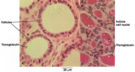
A. H. & E.; B. periodic acid-Schiff and hematoxylin stains, 612 x.
Follicles: Structural units of the thyroid gland supported and separated by connective tissue. Note variation in size. A single layer of cells forms the follicle. Shape of cells reflects functional activity. Cells in these follicles are cuboidal with central, rounded nuclei, indicating normal activity.
In A, the colloid in the lumen of the follicle is not stained. In B, the colloid is specifically stained red with the periodic acid-Schiff method because of the chemical composition of colloid, which is a glycoprotein-iodine complex (thyroglobulin).
Thyroid gland
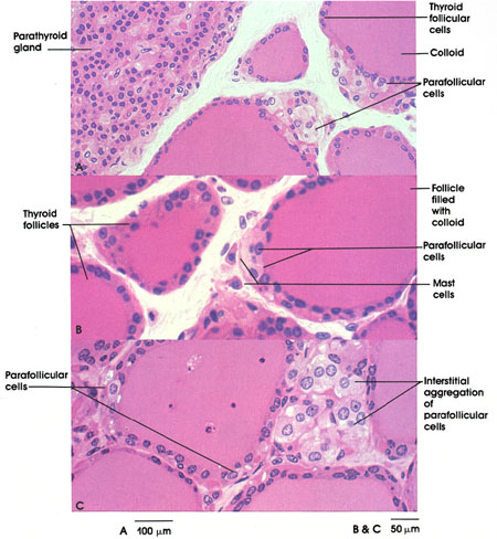
A. 119 x; B. & C. 169 x.
Parafollicular cells may be found intimately associated with thyroid follicles or as isolated or interstitial clusters of cells. They are not readily found in routine thicker sections of the thyroid gland. The photornicrographs above are from 1.5 µm plastic sections.
Although parafollicular cells (also known as light cells) appear, at the light microscopic level, to be in intimate contact with thyroid colloid, they are, in fact, separated from colloid by thin intervening processes of adjacent thyroid follicular cells.
Parafollicular cells synthesize and secrete the hormone calcitonin. This hormone lowers blood calcium levels by inhibiting bone resorption. The secretion of calcitonin results from elevation of blood calcium concentration above normal levels.
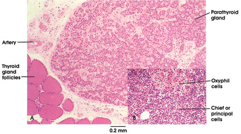
B. Human, 10% formalin, H. & E., 47 x.
Two cell types are found in the parathyroid gland. The most abundant type is the chief (or principal) cell, which secretes parathyroid hormone. Chief cells have a prominent nucleus, and a cytoplasm that stains variably and may be light or dark depending upon its secretory activity.
The second type, oxyphilic (acidophilic or eosinophilic) cells, occurs in small clumps and in fewer numbers. These cells usually have small densely staining heterochromatin and an oxyphilic cytoplasm whose perimeter is usually well defined. Oxyphilic cells usually increase in number with age but their specific function is unknown.
The parathyroids are essential for life. They control blood calcium and phosphate levels. A significant decrease in blood calcium results in tetany, abnormal twitching, and muscle spasms, caused by changes in excitability at the neuromuscular junction, and death.
Dietary addition of calcium and especially administration of parathyroid hormone relieves the abnormal spasms, preventing death of the organism.
Parathyroid hormone secretion is apparently regulated by blood calcium levels only in hypoparathyroidism, phosphate levels increase and calcium levels decrease. in hyperparathyroidism, blood phosphate is low and blood calcium levels are increased. Abnormal levels of calcium may result in abnormal deposition of calcium in the kidneys and muscle. Abnormally increased blood levels of calcium occur at the expense of bone, which may fracture as a result. Calcium removal from bone is related to osteoclastic activity, which is the site of action of parathyroid hormone.
The small parathyroid glands (4 to 5 mm in diameter) vary in number from three to six and are usually found on the posterior surface of the thyroid gland.
In the lower left corner, a few follicles of the thyroid gland can be seen.
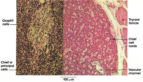
Parathyroid glands are essential for life. in the absence of parathyroid hormone, there is a pronounced decrease in blood calcium resulting in tetany, the intense, involuntary spasm of skeletal muscle.
Oxyphil cells: Occur in groups or nests among chief cells. Larger than chief cells, cytoplasm acidophilic. Oxyphils increase with age and are not found in all mammals. Function remains unknown.
Chief or principal cells: Much more numerous than oxyphil cells and functionally important. Nucleus round and centrally located. Cytoplasm homogeneous. Arranged in cords or plates separated by vascular channels. These cells produce parathyroid hormone, which is important in calcium metabolism.
Thyroid follicle: Seen adjacent to the capsule of the parathyroid gland
Islet of Langerhans
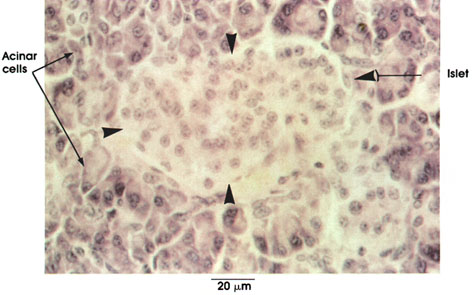
Islet: Pale-staining area demarcated by arrows. Contains irregular clumps of cells. Separated from the acinar cells by a thin partition of reticular tissue. This endocrine gland elaborates the hormones insulin and glucagon, which are secreted into the rich capillary bed and carried via the portal system of veins into and through the hepatic lobules before reaching the general circulation. Insulin and glucagon constitute an important system for the regulation of blood glucose levels. This endocrine organ is essential for life. Dysfunction results in diabetes mellitus, a common disorder of man, characterized in its uncontrolled and most severe form by polyuria (frequent urination), polydipsia (excessive water intake), hyperglycernia (elevated glucose level in blood), glycosuria (sugar in the urine), ketonuria (ketones in the urine), acidosis (inability to buffer the blood at pH 7.2 to 7.4), wasting of the body and, if not treated, coma and death.
Acinar cells: Darker staining, irregular cells. Rich in cytoplasmic ribonucleic acid (RNA). Elaborate digestive enzymes, which are carried to, and are active in, the duodenum. Pancreatic enzymes break down partially digested food from the stomach into simple compounds. Carbohydrates, fats, and proteins are hydrolyzed by the enzymes amylase, lipase, and various proteases (including the nucleoproteases) secreted by the acinar cells.
Islet of Langerhans
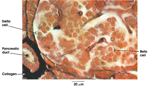
The islets of Langerhans were described by Paul Langerhans, a German physician, anatomist, and pathologist, in 1869. Although in routine histological preparations all of the islet cells appear to be similar, special methods reveal three types, alpha, beta, and delta.
Delta cell: Few in number compared to alpha or beta cells. Significance not well understood. Cytoplasm stains blue with Mallory-azan.
Beta cell: More numerous than alpha or delta cells. Produce insulin. Insulin increases cellular uptake of glucose and its conversion to glycogen. Beta cells may occur outside the islets. Granules are diffusely scattered in cytoplasm.
Pancreatic duct: Found interlobular connective tissue, lined by cuboidal to columnar epithelium. Size varies Collagen: In the interlobular connective tissue. Stains dark blue with Mallory-azan.
Alpha cells (not seen in this preparation) secrete the hormone glucagon, which effects the breakdown of liver glycogen and elevates the blood glucose level.
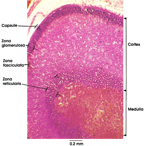
Capsule: A tough fibroelastic covering of the adrenal gland.
Cortex: Three concentric zones, each with a different cell arrangement, are shown. The adrenal cortex is essential to life. It controls the electrolyte and water distribution in the body and maintains proper carbohydrate balance.
Zona glomerulosa: Outermost narrow zone of the adrenal cortex. Deeply staining, densely packed nuclei. Elaborates aldosterone.
Zona fasciculata: The broadest zone of the adrenal cortex. Cells are arranged in long cords. Elaborates cortisol.
Zona reticularis: Innermost layer of adrenal cortex. Cells are arranged in irregular cords separated by sinusoids (clear spaces) giving the appearance of a meshwork. Stains deeper than zona fasciculata.
Medulla: Irregularly arranged mass of cells in cords. Highly vascularized. Rich in chrornaffin substance. Produces epinephrine and norepinephrine.
Chromaffin reaction
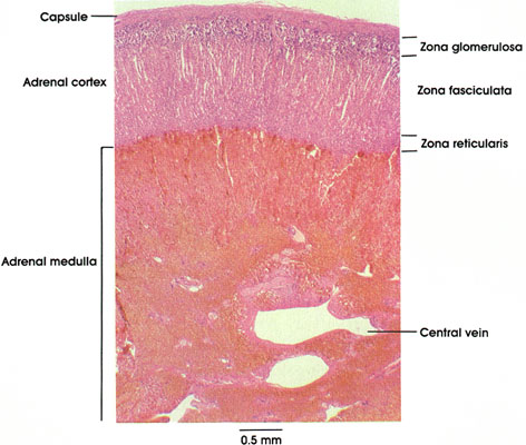
This low-power micrograph illustrates the structural organization of the suprarenal gland. There are two major subdivisions: cortex and medulla.
The suprarenal cortex exhibits three distinctly different structural layers: (1) zona glomerulosa, (2) zona fasciculata, and (3) zona reticularis.
The zona glomerulosa synthesizes and secretes mineralocorticoids, primarily aldosterone, which maintains sodium and potassium (electrolyte) and water balance. Zona fasciculata, and probably zona reticularis, synthesize and secrete cortisone, cortisol, and glucocorticoids, which are concerned with the regulation of carbohydrate, protein, and fat metabolism. Small amounts of androgen and estrogen are also secreted by zona fasciculata and zona reticularis. The cortical cells do not store their steroid hormones.
The medulla constitutes the core of the gland. In this preparation, the medulla has a distinctly brown coloration. Zenker's* fixative contains potassium dichromate, which oxidizes catecholamines to a brown (melanin-like) color. The affinity of adrenal medullary cells for chromium salts results in what is called a chrornaffin reaction;. the cells that produce the reaction are called chromaffin cells or chromaffin granule-containing cells. Chromaffin granules contain either epinephrine or norepinephrine, a binding protein, and dopamine B-hydroxylase.
ortex and medulla
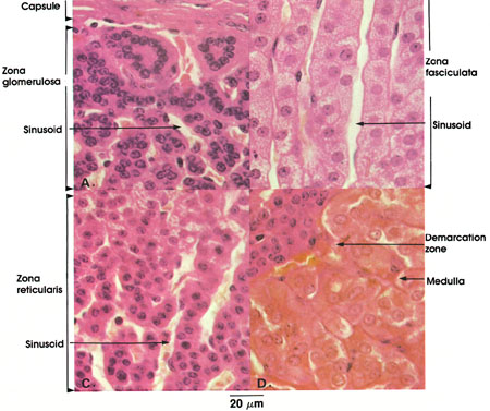
This plate illustrates the different zones of the adrenal gland, from the capsule to the medulla.
Capsule: A tough fibroelastic covering with delicate trabeculae extending into the substance of the gland.
Zona glomerulosa: Outermost narrow zone of the adrenal cortex. Cells arranged in ovoid groups without a significant lumen. Component cells are columnar with spherical, deeply staining nuclei. Richly supplied with blood. The zona glomerulosa secretes hormones concerned primarily with mineral metabolism. The mineralocorticoids are deoxycorticosterone and aldosterone.
Sinusoid: Sinusoids arise from multiple arterioles in the capsule. Course between cell cords.
Zona fasciculata: The middle and broadest zone of the adrenal cortex. Cells are regularly arranged in parallel cords, one to two cells thick. Component cells are cuboidal, frequently containing two vesicular nuclei. Vacuoles seen in some cells represent dissolved lipid droplets. Cholesterol is chiefly present in this zone. Cell cords are surrounded by sinusoids. Secretes the glucocorticoids, cortisone and cortisol.
Zona reticularis: Innermost layer of the adrenal cortex. Cells are arranged in irregular cords, are smaller than those of the zona fasciculata, and stain darker. Nuclei stain deeply. Sinusoids separate cell cords. Secretes the same glucocorticoids as the zona fasciculata. These hormones participate in carbohydrate, protein, and fat metabolism.
Demarcation zone: Shows zone of transition from zona reticularis to medulla.
Medulla: Polyhedral cells arranged in anastomosing cords. Prominent nuclei. Contain chromaffin granules, precursors of epinephrine and norepinephrine. Richly supplied with blood.
The adrenal cortex is essential for life, and, through its hormones, is involved in numerous body functions. These activities include the maintenance of water and electrolyte balance, carbohydrate metabolism, and the normal functioning of connective tissue cells. Destruction or removal of the cortex results in Addison's* disease unless cortical hormones are given to the patient.
The adrenal medulla is not essential for life. The hormones of the adrenal medulla influence the metabolic rate and cardiovascular function and induce lipolysis and the release of fatty acids from adipose tissue, and opiate-like pepticles, including leu- and met-enkephalin. Medullary cells store their hormones in the form of granules.
During normal activity only a small amount of catecholamine is secreted, but this is greatly increased during stressful periods of intense emotional reaction. It is believed that epinephrine and norepinephrine are secreted by two different types of cells. The epinephrine secreting cells are primarily located around the distal ends of sinusoids draining the adrenal cortex, whereas norepinephrine-secreting cells are associated with capillaries of the direct arterial supply of the medulla. All adrenal medullary cells are innervated by cholinergic nerve terminals of preganglionic sympathetic neurons.
Chromaffin cells are also found in paraganglia, which are collections of catecholamine-secreting cells associated with autonomic ganglia.
Heart coronary sulcus
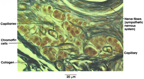
Scattered islands of chromaffin cells are found in the subepicardial connective tissue of the coronary sinus. Note relation to capillaries into which it is believed they pour their secretion of catecholamine. Chromaffin cells are usually closely associated with sympathetic nerve fibers and ganglion cells. The brownish coloration is due to oxidation of chromaffin granules by potassium dichromate in the fixative used in this preparation. Because of their structural similarity to cells of the adrenal medulla, it is assumed that chromaffin cell secretion augments action of the sympathetic nervous system by elevating blood sugar, increasing heart rate, raising blood pressure, and generally preparing the organism for emergency situations ("flight or fight").
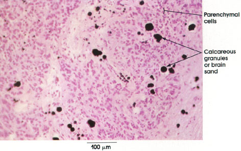
The pineal gland is made up of plates of cells separated by septa. Two cell types are recognized by special techniques, the more common parenchymal cell and the neuroglial (astrocyte-like) supporting cell. The latter is found between clusters of parenchymal cells. The two cell types cannot be distinguished in ordinary H. & E. preparations. Concretions or brain sand characterize the pineal gland and increase with age. They have a mineralized organic matrix and, at high magnification, appear lamellated.