Section 2: Epithelial Tissue
The layer of cells that covers the outer, and lines the inner, body surfaces is designated as epithelium. In general, many of these cells have a free surface, which is actually or potentially exposed to the external environment (skin, and the respiratory tract), or to a moist environment continuous with the external environment (digestive, reproductive, and urinary tracts). Other epithelial cells, comprising glands found in underlying connective tissue, are in continuity with the surface epithelium by epithelial duct cells. The glandular epithelium secretes diverse products, which are carried to the external surface. The products of these glands include sweat, bile, urine, reproductive cells and associated glandular secretions, mucus, milk, digestive enzymes, hydrochloric acid, and so on. Some epithelial cells have migrated away and have lost contact with the free surface. These cells form distinctive cellular masses, which are termed endocrine glands. The secretory products of these cellular masses are delivered into the vascular system to be carried to their specific sites of activity by the blood stream. The endocrine system will be considered in Section 15 of this atlas.
It is important to remember that everything that enters or leaves the body is either modified or synthesized by epithelial cells or has diffused or has been transported through this tissue. The various functions of epithelium include protection, secretion, excretion, digestion, absorption, lubrication, sensory reception, and reproduction. Such a diversity of functional activity depends upon structurally diverse cell types and cell groupings.
Epithelia are classified by histologists according to cell layering and cell shape. On this basis, three distinct types of epithelium are recognized: (1) simple, which is a single cell layer; (2) pseudostratified, which is a single cell layer but appears to have two or more layers; and (3) stratified, which is composed of several to many cell layers. Only the simple and stratified epithelia have important subgroupings, which are classified according to the shape of the cells that are exposed to the free surface. The simple epithelia are described as squamous (sheets of flattened cells), cuboidal and columnar (in which the cells are greater in height than width when seen in most sections-these, too, are actually five- or six-sided in cross section). The stratified epithelia include stratified squamous, in which the superficial cells on the free surface are flattened; stratified cuboidal, in which the superficial cells on the free surface are cuboidal; and stratified columnar, in which the superficial cells on the free surface are columnar.
From a functional point of view, the simple epithelia carry out the most diverse activities, which include absorption, excretion, synthesis, secretion, and sensory reception, whereas the stratified epithelia have protective functions, serve as conduits or ducts, and produce reproductive cells. In order to serve their distinctive functional roles, epithelial cells often display distinctive cell membrane or surface modifications and appendages.
The epithelial types shown in this section represent the morphological varieties of simple and stratified epithelia. The structural features of many other epithelial cell types and groupings are found in other sections of this atlas, where their functional role will be considered in the context of organ function.
A classification of epithelial cell types and some of their locations in the body follows.
-
Simple Epithelium
-
Squamous
Innermost lining of blood and lymph vessels and the heart (endothelium). Lining of the pleural, cardiac, and abdominal cavities. Initial segments of ducts of glands. Air sacs or alveoli of the respiratory system. Renal glomeruli and corpuscles. Kidney tubules (thin segment of loop of Henle of the nephron). -
Cuboidal
"Germinal" epithelium covering the ovary. Ducts of many glands. Ciliary body of the eye. -
Columnar Stomach, intestines, and gallbladder of the digestive system. Small bronchi of the respiratory system.
Uterine tubes. The secretory cells of many glands (endocrine and exocrine) vary from cuboidal to columnar. Size and shape may vary with the functional state (e.g., thyroid gland). -
Pseuclostratified
Pharynx, trachea, and large bronchi. Male excurrent ducts (epididymis and vas deferens). Parts of the female and male urethra. -
Specialized
Glands of intestinal tract, nasal cavity, bronchi, uterine tubes, and accessory sex glands. -
Pigmented
Epithelium of retina. -
Neuroepithelium
Receptor cells of taste, hearing, and balance.
-
-
Stratified Epithelium
-
Stratified squamous
Keratinized and non-keratinized epithelium of skin, palpebral conjunctivum, oral cavity, esophagus, and anus. Urethra near the external orifice. Vagina. -
Stratified cuboidal
Ducts of sweat and sebaceous glands of the skin. Graafian follicles of ovary. -
Stratified columnar
Pharynx, larynx, urethra, and portions of the excretory ducts of salivary and mammary glands. -
Transitional (urothelium)
Renal calyces and pelvis, ureter, and urinary bladder.
-
Several specializations of epithelial cells found on their free or exposed surface include the brush or striated border of the absorbing cells of the intestine and kidney, motile cilia of the pseudostratified epithelium of the respiratory system, and non-motile stereocilia of the pseudostratified epithelium lining the epididymis. Specializations that structurally and functionally link adjacent cells together include the "terminal bars" illustrated in Plate and the "intercellular bridges" or clesmosomes associated with prickle cells found in stratified squamous epithelium
. Marked infoldings of the basal cell membrane, termed basal striations, are seen in certain active transport cells such as the proximal convoluted tubule cells of the kidney and ducts of certain glands
Between the basal surface of epithelial cells and the underlying connective tissue is the basement membrane, which varies markedly from place to place and in certain disease states. This extracellular structure has been shown by electron microscopy to have several components that are produced by both epithelial cells and the underlying connective tissue fibroblasts
CUBOIDAL EPITHELIUM
Brush border, basal striations proximal tubules kidney
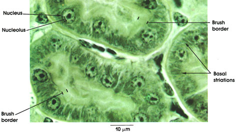
iron hematoxylin-orange G, 1416 x.
The cells of the proximal tubule with their apical and basal specializations have the capacity to reabsorb selectively and transport metabolically valuable substances from the glomerular filtrate (e.g., glucose and amino acids), returning them to the vascular system. They also transport and secrete other substances in the lumen of the proximal tubule to be eliminated in the urine.
Nucleus: Round and large with prominent nucleolus.
Brush border: On the luminal surface of the tubule cells. Consists of microvilli that vastly increase the cellular absorptive surface.
Basal striations: Consist of rod-shaped mitochondria contained within compartments formed by specialized infoldings of the basal cell membrane.
Kidney medulla collecting ducts
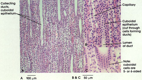
Human, 10% formalin, H. & E., A. 85 x; B. and C. 216 x.
The cells commonly referred to as cuboidal are always found to be five- or six-sided when cut through their axial or cross-sectional plane. This can be seen most readily in sections of the digestive system and kidney where almost every section contains both longitudinal and transverse profiles of cuboidal and columnar epithelia
UNICELLULAR GLAND
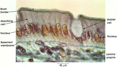
Cat, Helly's fluid, Mallory's stain, 1416 x.
Absorbing cell: Single cell layer of tall columnar cells. Basal ovoid nucleus. Although the cells appear retangular in this section, they are actually five- or six-sided when they are cross-sectioned. When the underlying tissue folds or bends, these cells may have a pyramidal appearance
Nucleus: Ovoid. Situated in lower half of the columnar cell. The nuclei in tightly packed cells may appear elongated and staggered at different levels within the cell. This is readily seen in pseudostratified ciliated columnar
Brush border: Also know as the straited border. Made up of fine, closely packed microvilli that vastly increase the surface area of the cell. Characteristic of absorptive surfaces. Adequate absorption of digestive products is dependent upon this cell surface specialization of absorbing columnar epithelial cells.
Goblet cell: Unicellular mucous glands scattered among the tall columnar cells appear empty because mucin is extracted during tissue processing. These unicellular gland cells are a specialization of simple epithelium and serve a protective function for the principal epithelial cell type.
Basement membrane: Delicate in appearance but a firm support for the columnar cells
Lamina propria: Connective tissue stroma. Reticular framework containing a.variety of wandering cells as well as vascular and lymphatic channels. Cells commonly found in the lamina propria include lymphocytes, plasma cells, eosinophils, and mast cells.
WITH STEREOCILIA
Epididymal duct
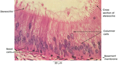
Rhesus monkey, Helly's fluid, H. & E., 612 x.
The epididymal duct is lined by a pseudostratified columnar epithelium containing two types of cells: tall columnar cells bearing so-called stereocilia and rounded basal cells. The cells forming a pseudostratified epithelium deceptively appear to be stratified in two or more layers. The cells actually vary in height, but all are in contact with the basement membrane.
Columnar cells: Tall cells bearing stereocilia. These are non-motile processes of the columnar cells projecting into the lumen. Although they are called cilia, electron micrographs show that they lack the structural characteristics of cilia, and they resemble greatly elongated microvilli. In this figure, they are seen in both cross and longitudinal section. Nuclei of columnar cells are elongated and lie at different levels.
Basal cells: Rounded or triangular cells, lying against the basement membrane, form a discontinous layer around the duct.
Pseudostratified columnar ciliated epithelium and
goblet cells trachea
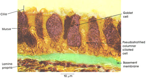
Rhesus monkey, Helly's fluid,
modified aldehyde fuchsin stain, 1416 x.
Epithelium: Pseudostratified ciliated columnar epithelium. The term pseudostratified refers to the appearance of the epithelium in section. Although the cells appear to be stratified because the nuclei are found in several layers, the basal portions of all cells are actually in contact with the basement membrane.
Cilia: These motile structures carry a carpet of mucus, provided by goblet cells, which collects inhaled debris and takes it to the pharynx where it is either coughed out or swallowed.
Goblet cells: These non-ciliated mucus-secreting cells are seen in various stages of mucous synthesis and discharge.
Basement membrane: This common structure is thickest in the trachea, but wandering cells of the immune system can be found traversing the membrane. Other cells of the immune system are also seen at various levels of the epithelium.
Lamina propria: The lamina propria of the trachea is thin but contains small blood vessels and collagenous and elastic fibers.
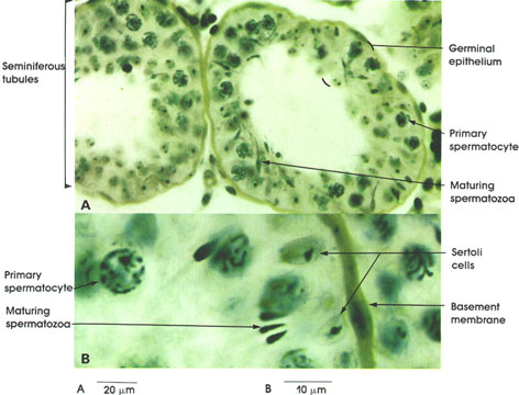
Rhesus monkey, Helly's fluid,
iron hematoxylin and orange G stains,
A. 162 x; B. 1416 x.
The germinal epithelium of the seminiferous tubules is composed of several layers of spermatogenic cells disposed between the basement membrane of the tubule and the lumen
Primary spermatocyte: Largest germ cell. Nuclei are large and vesicular and have condensed chromatin. Chromatin may appear as elongated threads.
Maturing spermatozoa: Mature germinal cell consisting of a head and a tail. The heads are in close association with Sertoli* cells, and the tails project into the lumen of the seminiferous tubule. Condensed nuclei forming the heads of spermatozoa contain a single set of chromosomes. Spermatozoa are the source of testicular hyaluronidase, an enzyme that may play a role in fertilization.
Sertoli Cell: These are supporting cells of the testicular epithelium. Tall columnar cells extend from the basement membrane to the lumen. These cells possess ovoid nuclei with a prominent nucleolus (seen here). Cell borders are not distinguished with light microscopy. Spermatozoa develop in intimate relation with the apical cytoplasmic processes of Sertoli cells.
Basement membrane: Surrounds seminiferous tubules, and is augmented by outer layers of connective tissue.
A. Non-keratinized B. Keratinized
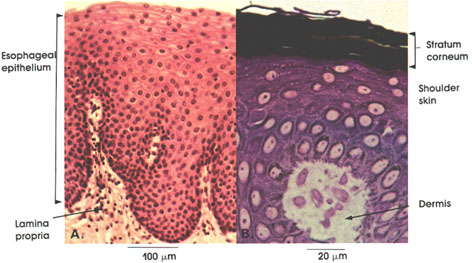
A. Human, 10% formalin, H. & E., 162 x.
B. Human, glutaraldehyde-osmium fixation, toluldine blue stain, 612 x.
Stratified squamous epithelium is made up of several layers of cells. The deepest layer is composed of cuboidal or low columnar cells, the middle layer of polygonal cells, and the superficial layers of flattened cells. The epithelium caps connective tissue papillae (lamina propria or dermis). The stratified squamous epithelium located internally (esophagus) is non-keratinized, whereas that located externally (skin) is keratinized (i.e., possesses a stratum corneum). The stratum corneum is made up of flattened non-viable, non-nucleated epithelial cells containing keratin
Mucous gland duct tongue
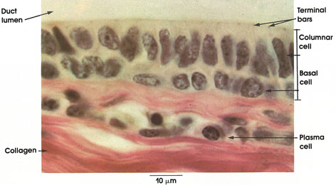
Human, Zenker's fluid,
iron hematoxylin & carmine stain, 1416 x.
Columnar cell: Columnar cells form the superficial layer of the stratified columnar epithelium.
Basal cell: These cells are irregularly polyhedral and form the deep layers of this stratified epithelium.
Terminal bars: Darkly stained, thickened zone is the specific attachment site of the lateral surface of adjacent superficial columnar cells (so-called junctional complex from electron microscopic studies).
Collagen: A component of the connective tissue stroma.
Plasma cell: Eccentrically placed, prominent and structurally characteristic nucleus in an abundant basophilic cytoplasm. Plasma cells produce antibodies
Ureter
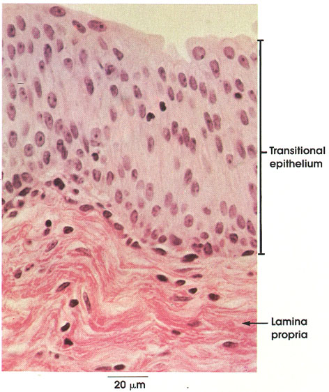
Human, Helly's fluid, H. & E., 612 x.
The term transitional epithelium does not imply that this epithelium is in actual transition from one type to another, but rather refers to the appearance of the cells, which changes as the organs with which they are associated are stretched or relaxed.
Transitional epithelium (uroepithellum): This stratified epithelium is found lining the urinary tract from the renal calyces to the urethra. It is in direct continuity with the simple epithelium of the ducts and collecting tubules of the kidney and the stratified squamous epithelium of the urethra. Superficial cells are cuboidal and large, and the basal cells are cuboidal to columnar. The surface cells of this epithelium vary in shape from squamous when stretched to columnar when contracted. Note the convex luminal border of the surface cells. These cells may be multinucleated and polyploid.
Lamina propria: Predominantly reticular and collagenous connective tissue fibers with some elastic fibers. The lamina propria contains many cells, including lymphocytes, plasma cells, eosinophils, and mast cells, in addition to blood capillaries and lymphatic vessels.
PIGMENT EPITHELIUM
Eye choroid layer
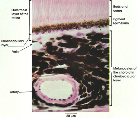
Rhesus monkey, Helly's fluid, H. & E., 612 x.
The choroid layer of the eye is a highly vascular and pigmented coat surrounding the retina. Shown in this figure is a part of the retina adjoining the choroid layer, as well as the major choroid layers. In the outermost layer of the retina, the following structures are seen:
Rods and cones: Neuroepithelial cells sensitive to light, arranged vertically and parallel.
Pigment epithelium: Single layer of pigmented cuboidal epithelial cells firmly bound to the choroid layer. Contains melanin pigment. In retinal detachments, the pigment epithelium remains attached to the choroid. The two major layers of the choroid seen in this plate are the following:
Choriocapillary layer: Composed of a network of wide lumen capillaries disposed in one plane and separated by delicate connective tissue fibers. Note that pigmented cells are essentially lacking in this layer. This layer supplies nutrition to the cells of the outermost layers of the retina.
Choriovascular layer: Filled with pigmented cells (melanin) and large-sized vessels
GLANDULAR EPITHELIUM
Zymogen pancreatic acinar cells
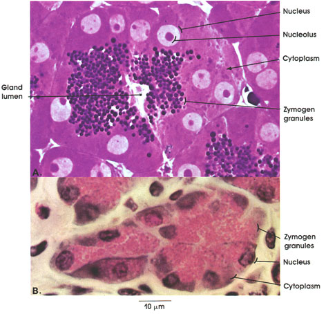
Rhesus monkey;
A. Glutaraldehyde-osmium fixation; toluldine blue and periodic acid-Schiff* stains;
B. Helly's fluid, Gomori's chrome alum hematoxylin; 1416 x.
The configuration of pancreatic acinar cells is seen in these two preparations.
Note the pyramidal shape of the acinar cell, the basally located round nuclei with distinct nucleoli, and two discrete zones of the cytoplasm. The apical zone near the lumen contains zymogen granules; the basal zone is intensely basophilic and free of granules. Electron microscopy has shown that the intense basophilia of the basal zone is due to its rich content of ribonucleoprotein bound to membrane (endoplasmic reticulum). Protein synthesized by the rough encloplasmic reticulum is transported to the supranuclear Golgi apparatus where zymogen (secretory) granules are formed, which are in turn transported to the apical cytoplasm, where they are discharged by exocytosis into the acinar lumen