Section 6: Nervous Tissue
The nervous system is developed entirely from ectoderm. It manifests optimally the two properties of protoplasm, irritability and conductivity, and is one of the most highly differentiated tissues in the body.
Neural tissue is made up of cells and their processes. Cells of the nervous system fall into two general categories: (1) nerve cells or neurons, and (2) supporting and satellite cells. In addition, neural tissue contains blood vessels and protective coverings (meninges).
The study of neural tissue is facilitated by several stains, none of which alone is capable of revealing all the desired details of structure. Because of the affinity of nerve cells and their processes for silver solutions (argyrophilia), silver impregnation methods are frequently used to demonstrate them.
The Golgi silver methods selectively impregnate relatively few cells, but they accomplish this most completely. These methods are good for outlining the external shape of nerve cells and their processes (especially dendrites) but do not reveal details of internal cell structure such as neurofibrils and Nissl bodies.
The Cajal and Bielschowsky silver methods are later developments of the silver impregnation methods. They are used to demonstrate axons, neurofibrils, and nerve endings, including synapses. Originally used on blocks of tissues, they have been modified for use on mounted sections. The most useful modification is that of Bodian in which activated protargol (silver proteinate) is used ).
The Nissl substance (cytoplasmic ribonucleoprotein) of nerve cells is revealed using basic aniline (cationic) dyes, also called Nissl stains, such as cresyl violet, gallocyanin, and toluidine blue (which bind to nucleic acid and demonstrate nuclei, nucleoli, and cytoplasmic Nissl substance of neurons.
Demonstration of the myelin sheath is accomplished by a variety of methods, including osmium tetroxide, Pal-Weigert, Weil, and Marchi techniques. The Marchi method is used to demonstrate degenerating myelin whereas the Pal-Weigert and Weil methods stain normal myelin ).
A neuron (nerve cell) consists of the cell body (perikaryon) and all its processes. Neurons ranging in diameter from 4 to 135 µm are generally larger than other cells in the body. The shape of neurons varies with the number and arrangement of their processes. In general, three types of neurons are recognized: (1) Unipolar or pseudounipolar neurons have spherical cell bodies with single processes that later bifurcate. Such cells are found in the dorsal root ganglia (2) Bipolar neurons are spindle-shaped, with one process at each end. Such neurons are found in certain peripheral ganglia, such as in the acoustic and olfactory systems .(3) Multipolar neurons have polygonal cell bodies and many processes. Such neurons are encountered in the autonomic ganglia and central nervous system ).
Nuclei of neurons are usually large, rounded, and centrally located and are characterized by well- defined, strongly RNA-positive nucleoli .Bi- and trinucleated neurons are found rarely in some autonomic ganglia).
The cytoplasm of neurons is rich in Nissl bodies, which are particularly coarse in the somatic motor neurons .Electron microscopy has shown the Nissl substance to be composed of ribonucleoprotein bound to membrane (granular encloplasmic reticulum). Nissl material extends into the proximal portions of dendrites but is absent from axons and axon hillocks. Nissl substance undergoes definite changes in response to axonal injury. In addition to the Nissl substance, neuronal cytoplasm is rich in mitochondria and contains a prominent perinuclear Golgi apparatus .The Golgi area of the neuron is the site where carbohydrates are linked to proteins in the synthesis of glycoproteins. Neurofibrils are seen in the cytoplasm of neurons and their processes.They are made up of subunits (neurofilaments), which are 7.5 to 10 nm in thickness and thus beyond the limit of resolution of the light microscope. Neurofilaments are made of structural proteins similar to those of the intermediate filaments of other types of cells. The argyrophilic neurofibrils are unique to nerve cells. In addition to neurofilaments, neuronal cytoplasm contains microtubules similar in external diameter (about 25 nm) to those observed in other types of cells. They are involved in the rapid transport of protein molecules through axons and dendrites. In addition to the aforementioned cell organelles, neuronal cytoplasm may contain lipid droplets, glycogen, pigment granules and secretory products .Pigment granules increase in number with age. Some types of neurons, such as Purkinje cells of the cerebellum, do not contain pigment granules.
Neurons in the central nervous system have a variety of shapes. They may be stellate in the anterior horn of the spinal cord or flask-shaped, as in the Purkinje cells of the cerebellum.Neurons in the peripheral ganglia are surrounded by satellite cells forming a capsule around the neuron. Those located in sensory ganglia are unipolar (pseudounipolar), whereas those in autonomic ganglia are multipolar.
The cell body of a neuron is its trophic center. Separation of a process from the cell body results in the death of that process. Neuronal processes are extensions of the cell body and serve to initiate or conduct nerve impulses. Dendrites generally receive and then conduct impulses toward the cell body, whereas axons conduct them away from the cell body. In unipolar neurons, in which the single process bifurcates into a peripheral and a central branch, both branches are structurally axon-like. In bipolar neurons, the effector and receptor portions of the neuron are found at the extreme ends of the two processes, and the entire intermediate portion is conductive. Multipolar neurons have several dendrites arising from the cell body and one axon that arises from the cell body or from the base of a dendrite . Dendrites branch repeatedly, and their surfaces are studded with spines or gemmules thus expanding the receptive cell surface. It is estimated that some neurons receive as many as 100,000 axon terminals on their dendritic expansion. A striking example of the vast dendritic expansion is seen in Golgi preparations of the Purkinje cell of the cerebellum ).
Axons are more slender than dendrites and are more uniform in diameter. The region of origin of the axon from a nerve cell is termed the axon hillock and is devoid of Nissl substance. It is the most excitable part of the neuron and the site at which the nerve impulse is initiated. Distally, each axon breaks up into simple or complex arborizations, the telodendria, which end on other neurons, glands, or muscle .Axons invariably acquire sheaths along their course. The axon and its sheath are referred to as a nerve fiber. Nerve fibers that run together in a bundle and share a common origin and destination in the central nervous system constitute a tract .A nerve fiber bundle in the peripheral nervous system constitutes a nerve.Nerve fibers may be myelinated or unmyelinated. Myelin sheaths are elaborated and maintained by oligodendroglia in the central nervous system, and by Schwann cells in the peripheral nervous system. Unmyelinated and myelinated peripheral nerve fibers are in intimate contact with Schwann cell cytoplasm and nucleus (the neurolemmal sheath or sheath of Schwann), and the plasma membrane is covered by a prominent polysaccharide surface coat. The relationship of such nerve fibers to oligodendroglia in the central nervous system is not quite so intimate. The myelin sheath around an axon is interrupted at regular intervals known as the nodes of Ranvier .The nodes are the site of voltage-gated sodium channels and ionic movement of impulse conduction. The flow of an electrical impulse along the nerve fiber thus skips from one node of Ranvier to the next. Myelin sheaths serve to insulate axons between nodes and thus speed up conduction of the nerve impulse between nodes of Ranvier (saltatory conduction). Myelin is made up of a lipid-protein complex. Some of the lipid is usually lost during tissue preparation, leaving behind a resistant proteo-lipid, neurokeratin unless special methods are used to preserve it ).
In addition to the myelin sheath and the sheath of Schwann, peripheral nerve fibers are surrounded by connective tissue, the endoneurium. The endoneurium is continuous with the more abundant connective tissue perineurium, which envelops bundles of nerve fibers. The nerve trunk is ensheathed in turn by the epineurium.
Nerve fibers, both axons and myelin sheaths, vary in size. The size of the nerve fiber (axon and its myelin coat) bears a direct relationship to its rate of impulse conduction. Large and heavily myelinated fibers conduct faster than small, unmyelinated ones.
Axons that branch at their termination to establish synapses on other neurons (dendrites, perikarya, or other axons) or muscle come in close proximity to, but not in contact with, the post-synaptic components of the synapse. Synaptic junctions vary in configuration, from the bouton-type (end bulb) of synapse , to the side-to-side contact seen in the climbing fiber system of the cerebellum (Plate 95), to the basket-type seen in the cerebellum ).
Supporting cells of the nervous system include the capsule or satellite cells of peripheral ganglia, ependyma, neuroglia, and Schwann cells.
Satellite cells surround neurons of peripheral ganglia, forming a capsule one cell layer thick . They are derived from neural crest elements and are continuous with a neurolemmal (Schwann) sheath.
Ependymal cells line the cavities of the brain and spinal cord A specialized form of ependymal cell is seen in some areas of the nervous system (subcommissural organ).
Neuroglia are the "supporting elements" of the central nervous system. Three cell types are found: (1) astrocytes with their two varieties, protoplasmic and fibrous; (2) oligodendroglia; and (3) microglia.
The astrocytes, as their name implies, are star-shaped cells with relatively lightly staining nuclei and processes closely applied to capillary blood vessels (perivascular end-feet or footplates). Other end- feet are applied to the pia mater. Two varieties are distinguished on the basis of the morphology of their processes. The protoplasmic variety, found mostly in gray matter, have plump and abundant cell processes that branch repeatedly .The fibrous variety, found mostly in white matter, have more slender but well-defined and fewer cell processes. They are longer and straighter than are those of the protoplasmic variety .Both varieties of astrocyte play a role in metabolite transfer within the central nervous system. The fibrous astrocytes, in addition, play a role in healing and scar formation in the nervous system. The cytoplasm of astrocytes contains glial filaments made up of glial fibrillary acidic protein (GFAP). Special histochemical stains for GFAP help identify astrocytes in tissue sections.
Oligodendroglia are smaller than astrocytes and have a denser nucleus and cytoplasm. As their name indicates, they have few delicate processes .These glial cells are seen adjacent to myelinated nerve fibers in the white matter or forming satellite cells to the neurons in the gray matter. Oligodendroglia elaborate central nervous system myelin.
Microglia are the smallest of the neuroglia, and, unlike the ectodermally derived macroglia (astrocytes and oligodendroglia), they are formed from the mesoderm. They are dense cells with deeply staining elongated nuclei and are frequently seen in gray matter in close proximity to neurons. The perikaryon of a microglial cell is irregular in shape, and, if elongated, the few processes emanate from both of its poles. Microglia are believed to be the scavenger cells of the central nervous system.
The central nervous system is covered by three protective coats (meninges): (1) The outermost layer is the dura mater, made up of a vascular dense fibrous connective tissue. (2) The middle layer is the arachnoid, a non-vascular delicate connective tissue coat. (3) The innermost layer is the pia mater, a delicate vascular layer adherent to the surface of the brain and spinal cord. Between the pia and arachnoid membranes is the subarachnoid space, in which the cerebrospinal fluid circulates. The small arteries and capillaries of the pia mater in certain regions of the ventricular system form tufts, which invaginate into the ventricular cavity (choroid plexus). The invaginated tufts are lined by cuboidal epithelium. The chorold plexus elaborates cerebrospinal fluid.
Peripherally located receptors constantly feed information into the central nervous system. These receptors may convey general sensation such as touch, pain, thermal sense, pressure, position, and movement or specialized sensations such as vision, audition, taste, and smell. The latter variety are discussed in Section 16. Illustrations of most of the former are seen in this section. Such receptors are found as (1) free nerve endings in epithelia or connective tissue, or as (2) encapsulated endings, in which the neural component of the receptor is surrounded by a connective tissue sheath of varying thickness. The sheath is continuous with the endoneurium and perineurium of the nerve fiber. Examples of such encapsulated endings are Meissner's (Krause's genital and Pacinian corpuscles the neuromuscular spindleand the Golgi tendon organ
Stimulation of any of these receptors results in the initiation of a nerve impulse that travels to the central nervous system. The translation of this impulse into a conscious sensation is a function of the brain.
Although doubt has been cast upon the functional specificity of the different varieties of receptors, it is still generally believed that free nerve endings respond optimally to sensations of pain, and possibly touch and thermal sense; Meissner's corpuscles respond optimally to touch sensations, whereas Pacinian corpuscles respond optimally to pressure sensibility. Krause's end-bulb and Ruffini receptors are believed to be cold and warmth receptors, respectively. The receptors in muscle and tendon are concerned with movement and posture. They respond to stretch and tension resulting from muscular contraction or passive stretch of muscles.
Postcentral gyrus cell layering
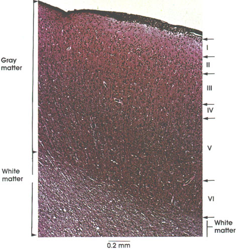
Lamination is a major characteristic of cortical structure. Six horizontal laminae distinguish the neocortex. Laminae are differentiated by the type, density, and arrangement of cells. The six laminae seen in this plate are, from the surface of the cortex to the white matter, as follows.
I-Molecular layer or plexiform layer: Contains few cells and a rich nerve fiber plexus made up of axons and dendrites of cells in other laminae as well as cells in this lamina.
II-External granular layer: Closely packed small neurons.
III-External pyramidal layer: Composed mainly of pyramidal neurons and many granule cells and cells of Martinotti.*
IV-Internal granular layer: Composed chiefly of stellate cells that are closely packed.
V-Internal pyramidal or ganglionic layer: Consists of medium-sized and large pyramidal cells intermingled with granule cells.
VI-Multiform layer or layer of fusiform cells: Contains a variety of cell types.
White matter: Contains incoming and outgoing nerve fibers.
Motor area, Precentral region

Pyramidal cells: Characteristic of cerebral cortex. The apex of the cell directed toward the surface of the cortex is termed the apical dendrite. Horizontally oriented basal dendrites also arise from the cell body. Note the variation in size of pyramidal cells (10 to 100 µm). The pyramidal cells located in the motor cortex are the largest of their kind and are known as Betz* cells. The region between pyramidal and glial cells is termed the neuropil and is filled with glial and nerve cell processes
Betz cell
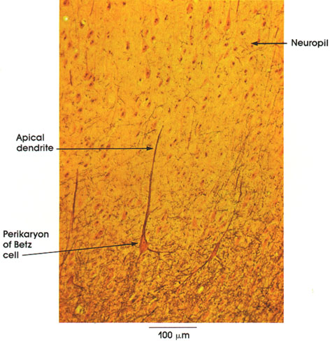
This is a section from the cerebral motor cortex (Area 4), where the pyramidal cells of Betz are found. Note the large multipolar cell body (perikaryon) and the apical dendrite directed toward the surface of the cortex. Surrounding these nerve cell bodies are processes of neural and glial origin (neuropil). In the neuropil, innumerable synaptic contacts (not seen by this method) occur between nerve cells and their processes.
Pyramidal cells
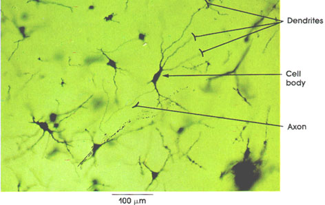
Cell body: Pyramidal in shape.
Dendrites: Each cell possesses several large tapering processes containing cytoplasmic organelles similar to those found in the cell body. Some dendrites possess spine-like side processes called gemmules,.
Axon: This process arises in this particular cell from the proximal part of the dendrite. It is slender and of uniform diameter, but variable in length depending upon location. The axon of a motor neuron may exceed 1 m in length.
Pyramidal cell
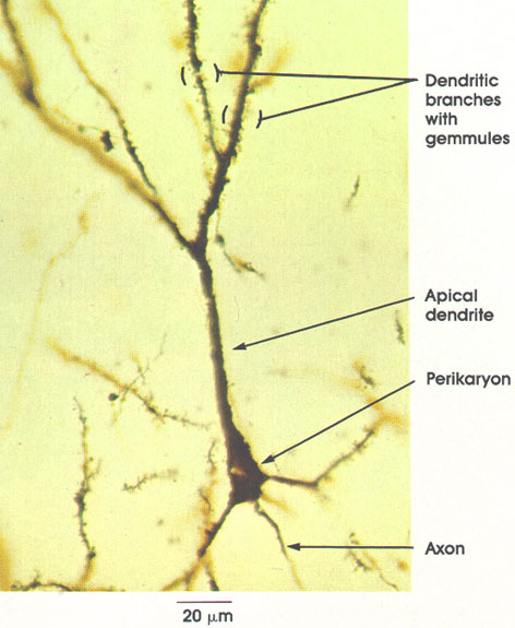
Perikaryon: Pyramid-shaped cell body impregnated with silver. Details of inner structure of nerve cells are not revealed by this method.
Apical dendrite: Stout tapering process. Directed toward surface of brain. Highly branched (Greek, dendron, tree).
Dendritic branches with gemmules: Dendritic branches increase the neuron surface for reception of many axon terminals or synapses. Dendritic branches are studded with spiny processes (gemmules) that increase greatly the surface area of the dendrite. It is estimated that this elaboration of surface area allows large neurons to receive as many as 100,000 separate axon terminals or synapses.
Axon: A single axon arises from the nerve cell body (as in this figure) or from the proximal part of a dendrite .It has slender distal extensions with a smoother contour than dendrites and a uniform diameter. The method used in this preparation is the only one capable of revealing the whole neuron (perikaryon and its processes).
Spinal cord ventral horn
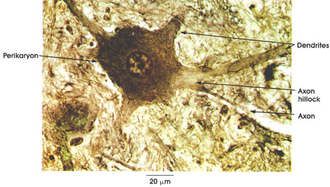
Perikaryon: It is multipolar (i.e., possesses a single axon and several dendrites) and has a central prominent nucleus. Cytoplasm is rich in Nissl* bodies except at the axon hillock (lighter area of cell body from which axon arises). .
Dendrites: Stout tapering processes similar in structure to the perikaryon.
Axon: This arises from the axon hillock. It is a slender process of uniform diameter and great length. The myelin sheath is not seen around the axon in this preparation because it is not preserved by the fixation method used.
Motor neurons Spinal cord
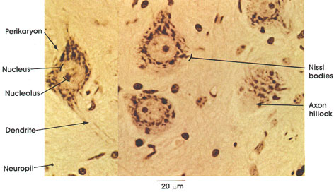
Perikaryon: Cell body, multipolar and large.
Nucleus: Spherical, pale, centrally placed with widely dispersed chromatin.
Nucleolus: Prominent in the pale background of the nucleus.
Dendrite: A process of the neuron that allows an expansion of the neuron surface for reception of stimuli. Nissl material is restricted to the proximal region of the dendrite.
Neuropil: Region between neurons. Composed of neuronal and glial processes.
Nissl bodies: Named after the German histologist Nissl, who first described them. They are one of the major characteristics of the neurons. Found in perikarya and in the proximal part of the dendrite. Electron microscopy reveals Nissl substance to be composed of ribosomes bound to membranes (the rough endoplasmic reticulum). Nissl substance is involved in protein synthesis. Nissl bodies undergo a distinctive change (chromatolysis) in response to axon section or injury.
Axon hillock: The region in the neuronal cell body that marks the emerging axon. It is devoid of Nissl bodies
Spinal cord, Lower motor neuron
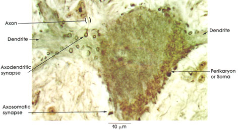
Axon: Preterminal portion before establishing contact.
Dendrite: Stout branching process.
Axodendritic synapse: Synaptic knob (also called bouton) arising from an axon that is in contact with a dendritic process. The synaptic knob or axon terminal swelling is actually a sac containing minute vesicles and mitochondria and not a loop or ring as seen in preparations of this type.
Axosomatic synapse: Axon terminal knob in contact with the cell body or perikaryon of the motor neuron.
Perikaryon or Soma: These are multipolar and are studded with terminal knobs. Variation of fine focus in such preparations will reveal the richness of synaptic terminals. The synaptic knobs seen in this photomicrograph represent only a minute fraction of the total synaptic terminals. It has been estimated that as many as 1200 to 1800 synaptic knobs may establish contact with one spinal motor neuron
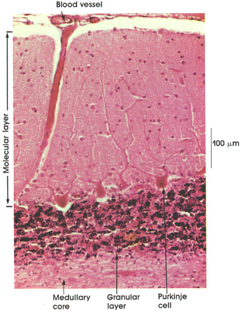
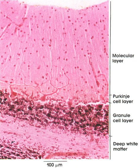
Medullary core: The white matter of the cerebellum contains nerve fibers that carry the afferent input and efferent output of the cerebellum.
Granular layer: Closely packed with the nuclei of small neurons (granule cells). Receives the major input to the cerebellum (the mossy fiber input).
Purkinje cell: Single row of large flask-shaped neurons. Dendrites arborize richly in the molecular layer. Axons exit in the medullary core but are not seen in this preparation.
Molecular layer: This is the most superficial layer. It is sparsely cellular and is largely a synaptic layer. It is primarily composed of the dendrites of Purkinje, stellate, Golgi, and basket cells and axons of granule cells.
Blood vessel: Located in the subarachnoid space, blood vessels penetrate deeply into folds of the cerebellum to nourish and remove metabolic waste products from the nerve cells and neuroglia..
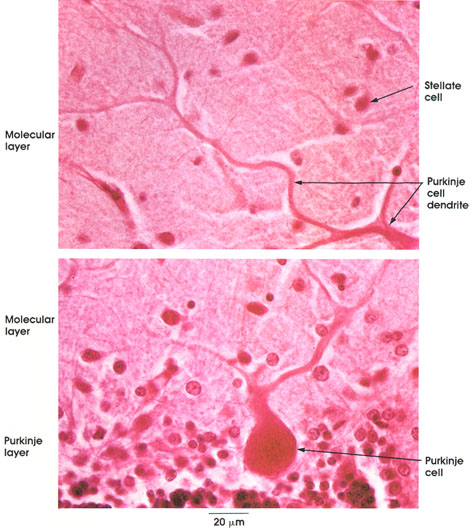
Molecular layer: Most superficial layer of the cerebellum. It is sparsely cellular and is largely synaptic.
Stellate cells: Sparsely scattered in the molecular layer. Usually small cells with short dendrites and fine unmyelinated axons that run horizontally. Larger stellate cells in the vicinity of Purkinje cells are known as basket cells.
Purkinje layer: Single row of large flask-like cell bodies situated between the molecular and granule cell layers.
Purkinje cell: These cells are flask-shaped. Each cell gives off two or three main dendrites, which arborize richly in the molecular layer. Their axons pass through the granule layer and enter the medullary core. They project upon deep cerebellar nuclei or on extracerebellar (vestibular) targets
20% formalin,
Bielschowsky's method, 612 x.
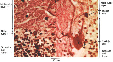
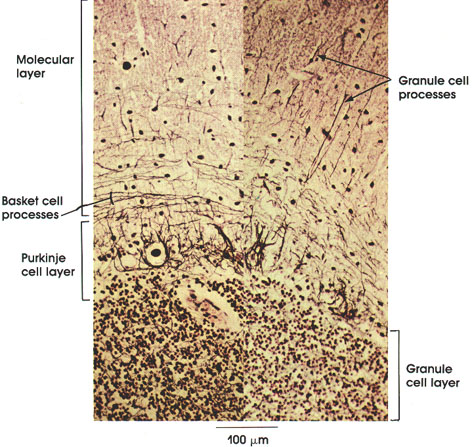
Molecular layer: Most superficial layer of the cerebellar cortex. It is sparsely cellular and largely synaptic.
Basket cell: A large variety of stellate cells deep in the molecular layer in the vicinity of Purkinje cells. Axons run transversely in the molecular layer and send collaterals that arborize around the perikarya of Purkinje cells like a basket.
Purkinje cell: Single row of large, flask-shaped cells. Form a distinct layer bordering the molecular and granule cell layers. Dendrites arborize richly in the molecular layer. These cells are named after Johannes Purkinje, a Bohemian physiologist, who described them in 1837.
Granule cell layer: Closely packed with chromatic nuclei of small granule cell neurons. Major input to the cerebellum projects into this layer.
Golgi Type II: This type of cerebellar neuron is found in the upper part of the granule cell layer close to the Purkinje cell layer. Larger than the granule cell neuron. Dendrites arborize extensively in the molecular layer. Axons establish synapses with dendrites of granule cells in the glomeruli of the granule cell layer. It is estimated that there is one Golgi Type II cell for every 10 Purkinje cells.
Cortex
Ranson's method,
612 x.
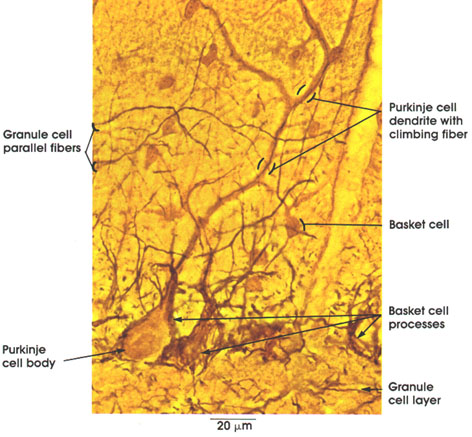
Granule cell parallel fibers: Axons of granule cells ascend from the granule cell layer to the molecular layer, where each divides into two branches that run horizontally across the layer, establishing synapses with dendrites of several Purkinje cells. They also establish synapses with Golgi, basket, and stellate cell dendrites.
Purkinje cell body: Large flask-shaped cell.
Purkinje cell dendrite with climbing fiber: Purkinje cell dendrites and climbing fibers run parallel to each other. The latter have an excitatory influence on Purkinje cells. The climbing fiber appears as a thin black line on the thicker brown-stained Purkinje cell dendrite.
Basket cell: A special variety of stellate cell in the molecular layer close to the Purkinje cells. Axons of basket cells run transversely in the molecular layer, giving off collaterals that form a "basket" arborization around Purkinje cells.
Granule cell layer: A layer of small granule cells under the Purkinje cell layer. it receives the major input to the cerebellum.
Ranson's method,
612 x.
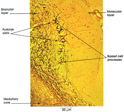
Molecular layer: Most superficial layer, containing few neurons. It is largely synaptic.
Basket cell processes: Axons of basket cells run transversely in the folium giving off collaterals that form a basket-like arborization around Purkinje cell bodies. They also synapse with Purkinje cell dendrites and proximal parts of the axon.
Medullary core: Deep white matter, which contains the entire afferent and efferent axons of the cerebellum.
Granule cell layer: A plexus or network composed of processes of granule cells and Golgi Type II cells as well as mossy fiber input. Mossy fibers are the terminations of all fibers entering the cerebellum except the olivocerebellar fibers, which constitute the climbing fibers.
Purkinje cells: Single row of large flask-shaped cells. Dendrites arborize richly in the molecular layer. Note black arborization of basket cell axons around the Purkinje cells.
Molecular layer: This is the most superficial layer. It is sparsely cellular and largely synaptic. Note dendrites of Purkinje cells arborizing in this layer..
Purkinje cells: Single row of flask-shaped large neurons. It has the largest neurons in the cerebellum. Dendrites arborize richly in molecular layer. Axons enter medullary core.
Granule cell layer: Closely packed with chromatic nuclei of small granule cell neurons. Lighter islands in this layer represent glomeruli where synapses are established.
Deep white matter: Contains afferent and efferent fibers of the cerebellum. Also known as medullary core.
Nerve cell processes
Molecular layer: Outermost layer of the cerebellum. Purkinje cell layer: Single row of large flask-shaped cells. .
Granule cell layer: Deep to the Purkinje cell layer. It contains small closely packed granule cells.
Granule cell processes: Axons of granule cells reach the molecular layer where they bifurcate (parallel fibers) to establish synapses.
Basket cell processes: Axons of basket cells, so-named because they form a basket-like arborization around Purkinje cells.
Purkinje cell
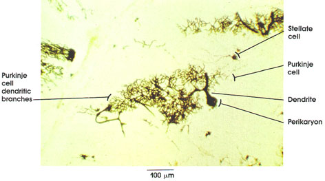
Purkinje cell: Single row of large cell bodies in the Purkinje cell layer of the cerebellum. Only one complete cell is seen in this figure.
Perikaryon: Flask-shaped Purkinje cell body impregnated with silver. Details of inner structure are not revealed by this method.
Dendrite: Directed toward surface of cerebellum. Each Purkinje cell possesses several main dendrites that enter the molecular layer.
Purkinje cell dendritic branches: Purkinje cell dendrites arborize richly in the molecular layer. The arborization is fan-shaped and extends at right angles to the cerebellar folia.
Stellate cell: Located in molecular layer. Small cell body with short, thin dendrites ramifying near the cell body, and fine unmyelinated axon extending transversely to the folia, which establishes synaptic contact with Purkinje cell dendrites.
Sensory neurons
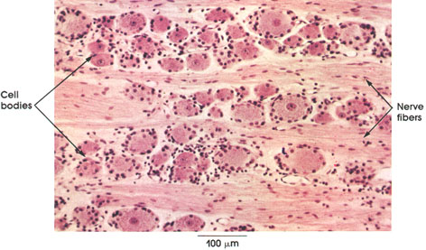
Cell bodies: Unipolar and ovoid or spherical in shape. Note the variation in size and density of staining of the large "clear" cells and the more numerous, small, densely staining "obscure" cells. . Each ganglion cell is surrounded by deeply stained nuclei of the capsule cells (satellite cells).
Nerve fibers: These are shown separating groups of nerve cells. They constitute central and peripheral axons of ganglion cells. Central and peripheral axons are branches of a single extension of the sensory neuron cell body .The peripheral processes convey impulses from sensory receptors, and, since they conduct toward the cell body, they function like a dendrite. The central processes carry impulses to the central nervous system (spinal cord). No synapses occur in the dorsal root ganglion.
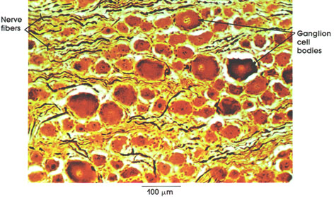
Ganglion cell bodies: Unipolar cells of variable size.
Nerve flbers: Myelinated fibers separate cell bodies. They constitute the afferent and efferent nerve fibers. In this preparation, the axons, but not the myelin sheaths, are stained. The myelin .
Cell bodies

Satellite cells: Of neural crest origin and concentrically arranged around ganglion cells. Also called capsule cells. Rounded or elongated nuclei are darker than nuclei of adjacent ganglion cells.
Perikaryon: Dorsal root ganglion cell body of neural crest origin. Ovoid or spherical in shape. Finely scattered cytoplasmic ribonucleoprotein (Nissl substance). Prominent central nucleus with a well- defined nucleolus. Indentations of the surface margin are caused by satellite cells.
Obscure cell body: Smaller and darker variety of ganglion cells. The nature and functional significance of obscure cells are uncertain.
Axons: Processes of ganglion cells.
Sensory neurons
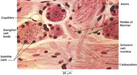
Ganglion cell body: This is ovoid or spherical. Note the variation in size and intensity of staining. The nucleus is central.
Satellite cells: Concentrically arranged around the ganglion cells.
Capillary: With enclothelial cell nucleus. Located in the connective tissue stroma between ganglion cells. Absence of blood cells from the capillary lumen is due to vascular perfusion fixation.
Axons: Processes of ganglion cells. Intricate coiling and winding denote its proximity to the cell of origin.
Nodes of Ranvier: Site of termination of myelin sheath segments.
Schwann cell nucleus: Flat to oval in shape. Close proximity to myelinated fibers. Schwann cells elaborate myelin sheaths.
T-bifurcation: Ganglion cell axon bifurcates to form peripheral and central processes; hence, dorsal root ganglion cells are termed pseudounipolar.
Nodes of Ranvier
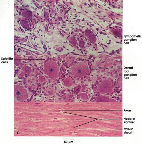
Compare and recognize the differences between (A) the smaller, oval, multipolar sympathetic ganglion cells with eccentric pale nuclei, and (B) the many significantly larger, round, pseudounipolar dorsal root ganglion cells with central nuclei. These ganglion cells are completely surrounded by a single row of small cells called satellite cells.
The sympathetic ganglion cells belong to the autonomic nervous system. They are motor neurons that, for example, innervate and activate smooth muscle of the vascular system and stimulate cells of the adrenal medulla to secrete their hormones, giving rise to an increase in blood pressure and heart rate. The dorsal root ganglion cells are strictly related to sensory receptors located in peripheral tissues, which provide an information link between external environmental conditions and the individual organism.
Nodes of Ranvier* are produced by the termination of the cytoplasm of two adjacent Schwann* cells, which wrap around axons forming myelin sheaths.
Cochlear nerve
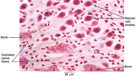
Bone: Modiolus or central conical pillar of spongy bone of the osseous cochlea.
Cochlear nerve fibers: Central processes of the bipolar ganglion cells.
Bipolar cell bodies: Spiral ganglion. Peripheral processes of sensory hair cells located in the organ of Corti.* Central processes from the spiral ganglion cells form the cochlear nerve (auditory part of the eighth cranial nerve).
Pseudounipolar neurons
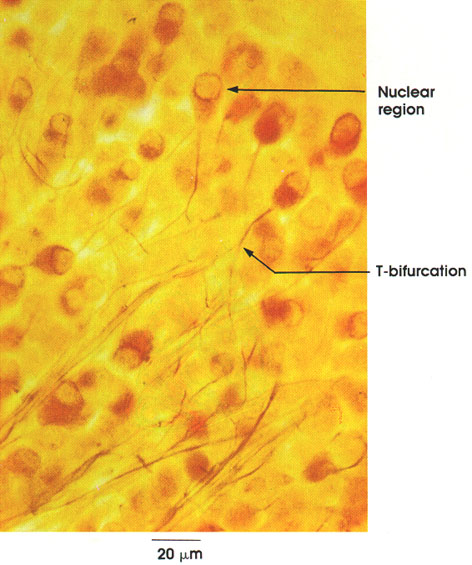
This plate is taken from an embryonic Gasserian* or trigerninal ganglion (fifth cranial nerve) and shows pseuclounipolar sensory neurons. Nuclei of neurons are seen as negative images. These neural processes characteristically bifurcate into central and peripheral extensions. The Gasserian ganglion is the "dorsal root ganglion" of the trigeminal nerve.
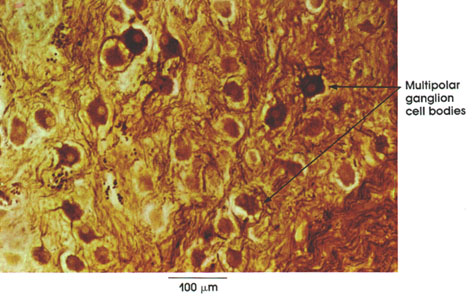
Multipolar ganglion cell bodies: Numerous dendrites, eccentric pale nucleus. Compare the multipolar ganglion cells of sympathetic ganglia with the unipolar ganglion cells of dorsal root ganglia and with the bipolar cells of sensory ganglia .Most neurons of the central nervous system are of the multipolar variety. Preganglionic nerve fibers from the thoracolumbar autonomic outflow of the spinal cord synapse with cells within sympathetic ganglia.
AND PARANEURONS
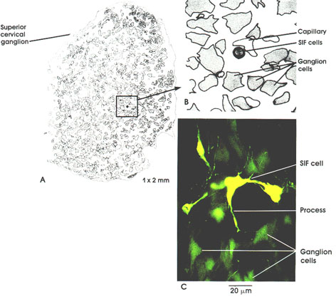
Adrenergic interneurons have been demonstrated in guinea pig sympathetic ganglia (by Jean Y. Jew, M.D.). Substantial surface area of these small intensely fluorescent (SIF) cells are in close relation to capillaries. As one can appreciate, the number of SIF cells in a ganglion is very small. However, these cells are reminiscent of enterochromaffin cells, widely, but also very sparsely distributed in the digestive tract. There is evidence suggesting that catecholamine transmitter leaves SIF cells as granules to enter blood vessels. This would permit transmitter originating in SIF cells to distribute widely to principal ganglionic neurons. Evidence of an intraganglionic portal system is based on the work of Drs. Christine Heym and Terence H. Williams.
Seminal vesicle
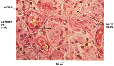
(Preganglionic fibers that synapse within parasympathetic ganglia are axons of neurons in the craniosacral division of the autonomic nervous system.
Ganglion cell body: Multipolar neurons. They have a large, eccentrically placed nucleus. Binucleate cells are commonly found in pelvic ganglia and occasionally in the heart. Note the dark nuclei of satellite or capsule cells surrounding the neuron.
Nerve fibers: Myelinated preganglionic and unmyelinated postganglionic fibers of the parasympathetic nervous system.
Venule: A venule is seen in the connective tissue stroma between ganglion cells. Blood is carried away in venules from the capillary bed supplying the ganglion cells and other tissue.
Pancreas
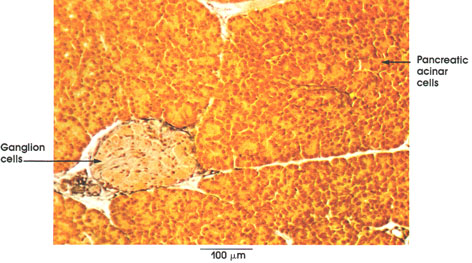
Pancreatic secretion is under neural and hormonal control. The chyme (semifluid mass of partially digested food mixed with gastric enzymes and hydrochloric acid) arriving in the intestine and contacting the intestinal epithelium results in pancreatic secretion. When hydrochloric acid and products of partial protein digestion (proteoses and peptones) contact the intestinal mucous membrane, two hormones are released and carried in the blood to the pancreas. The hormone secretin promotes the secretion of water and salts while the hormone pancreozymin depletes zymogen granules (digestive enzymes) from the pancreatic acinar cells. Zymogen granules are also secreted from acinar cells by vagal (parasympathetic) and splanchnic nerve (sympathetic) stimulation.
Secretin was discovered by Bayliss and Starling* in 1902. They correctly suggested that secretin was the first example of a whole group of chemical regulators (as yet to be discovered) produced in the body that could be designated as hormones.
Ganglion cells: Aggregates of parasympathetic ganglion cells enclosed in a thin connective tissue sheath between pancreatic lobules. Afferent input to these cells is from the vagus nerve.
Pancreatic acinar cells: Irregular clusters of pancreatic exocrine secretory cells arranged in lobules separated by thin connective tissue septa.
Myenteric and submucosal plexuses
A. ileum B. colon
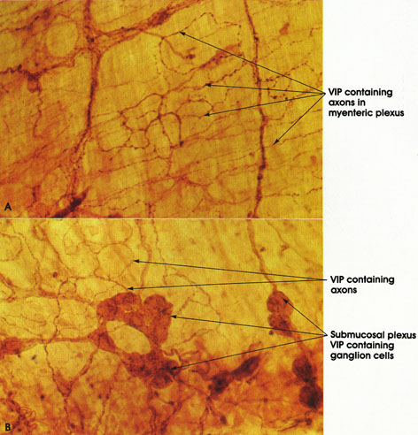
immunoperoxidase technique, A., B., 350 x.
The upper illustration (A) is of VIP containing nerve fibers of the myenteric plexus (of Auerbach*) as confirmed by the subtle cross-hatched appearance caused by the outer longitudinal and inner circular layers of smooth muscle fibers. The latter shows some background staining.
The lower illustration (B) is of VIP containing axons and ganglion cell bodies of the submucosal plexus (of Meissner*).
VIP is believed to increase intestinal motility.
Heart
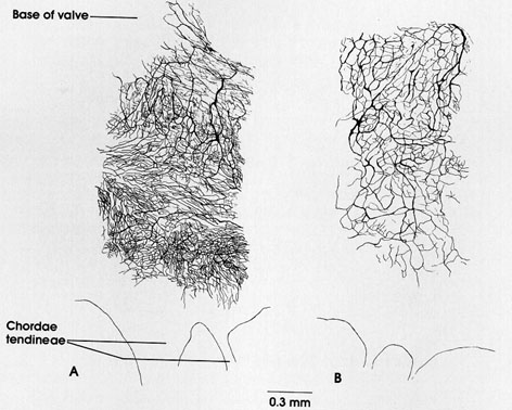
of Coupland and Holmes, A., B., 44 x.
This illustration demonstrates age-related decrease in autonomic nerve fiber bundles of the subendocardial region of the mitral valve. The cholinesterase-positive nerves shown here were drawn by camera lucida, which captures all the nerve processes stained at slightly different levels in the valve.
A is drawn from a young adult rat; B is drawn from a 3-year-old rat.
White matter, Spinal cord cross section
Axon: Shrunken axis cylinder appears black because of silver impregnation. Note the variation in axon diameter. The diameter of a nerve fiber is directly related to the speed of nerve impulse transmission; larger fibers carry electrical impulses faster than smaller fibers.
Myelin sheath: Unstained by this method. it surrounds the axis cylinder. .
A. Dorsal root B. Ventral root
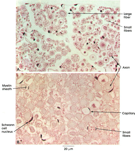
The method used here demonstrates axons but not myelin, hence the clear areas around the centrally placed axons represent unstained myelin. The partitions in the clear areas are artifacts of fixation.
Note that nerve fibers in the ventral root (B) are, on the whole, larger than those in the dorsal root (A). Note also the variation in fiber size within both the dorsal and ventral roots. Very small fibers seen in B are unmyelinated. The capillary seen in B shows a crescent-shaped dark endothelial cell nucleus and clear lumen (perfusion fixation). A large, dark, crescent-like Schwann cell nucleus is also seen in B. Schwann cells elaborate the myelin sheath. The dorsal roots are composed of the central processes of unipolar neurons in the dorsal root ganglion .The ventral roots are composed mainly of axons of the large somatic motoneurons, the smaller gamma motoneurons, and visceromotor axons from the autonomic intermediolateral cell column.
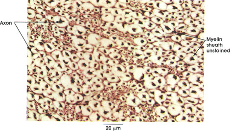
Sciatic nerve cross section
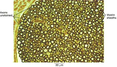
Axons unstained: Note variation in size.
Myelin sheath: Dense sleeve around axons. Myelin sheaths in the peripheral nervous system are elaborated by Schwann cells. .
Axon terminations of hypothalamic
secretory neurons
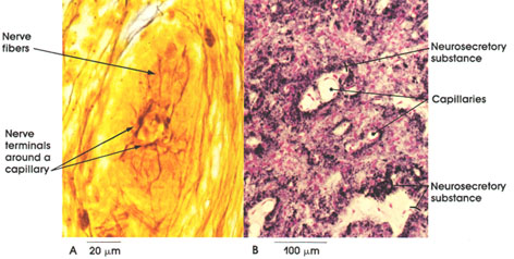
B. Rat, Helly's fluid, Gomori's chrom alum
hematoxylin and phloxine stains, 162 x.
The posterior lobe of the hypophysis, the neurohypophysis, is composed of nerve fibers that arise in the hypothalamus. Their cell bodies lie in the supraoptic and paraventricular nuclei. In A, nerve fibers and their pericapillary terminations are shown. In B, the method used selectively demonstrates neurosecretory material located in the axons. Note the abundance of blood vessels and the stained neurosecretory material surrounding the pericapillary spaces. It is believed that the nerve terminals release oxytocin and vasopressin into the blood stream. These two hormones are polypepticles. Oxytocin causes the smooth muscle fibers of the uterus to contract, a function essential for parturition, and can be used clinically to induce labor. Oxytocin also causes the myoepithelial cells of the mammary gland to contract, bringing about the flow of milk from the gland.
Vasopressin or antidiuretic hormone (ADH) raises blood pressure by acting on arterial blood vessels, stimulates the adrenal cortex, and increases the permeability of the distal and collecting tubules of the kidneys. This increases water reabsorption from the glomerular filtrate, inhibiting an abnormally large flow of urine (diuresis) and resulting in the formation of a urine hypertonic with respect to blood plasma
Lateral rectus muscle cross section
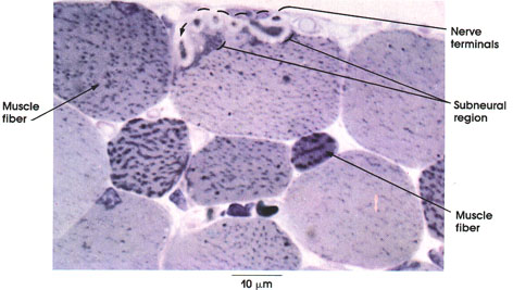
This is a 1 µm thin section of plastic-embedded tissue. Each method provides information not usually attainable by the use of only one method.
Nerve terminals: Nonmyelinated, dip into specialized sarcolemmal folds or gutters in the muscle fiber. The nerve fiber from which these terminals arise is not seen in this thin section.
Subneural region: Specialized synaptic region of the muscle fiber. Multiple sarcolemmal folds.
Muscle fiber: Note the variation in size of fibers and in staining pattern.
Intercostal muscle
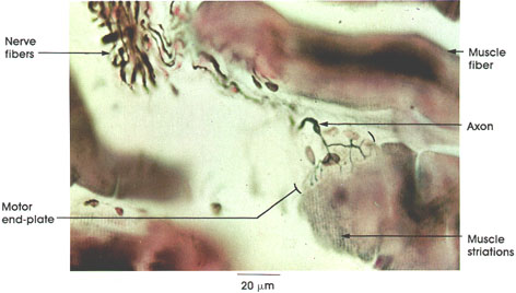
The tissue seen here was paraffin embedded and sectioned at 20 µm. This beautiful method permits a comprehensive view of the components of the nerve-muscle junction. The subneural .
Nerve fibers: Myelinated somatic motor nerve fibers branch extensively within and between the muscle fibers. Axons (but not their myelin sheaths) are stained in this preparation.
Axon: Although not shown, nerve fibers lose their myelin sheaths as they approach the motor end plate region. These nonmyelinated axons branch extensively on the surface of the muscle fiber.
Motor end plate: A well-defined junction of axon terminals on the muscle fiber surface. It is at this place that the electrical nerve impulse is chemically (acetylcholine) transferred to the muscle fiber. A muscle action potential is generated, and the electrical impulse is conducted over the fiber surface, resulting in muscular contraction.
Muscle striations: Note that the striated myofibrils do not extend inside the motor end plate region.
Muscle fiber: The number of muscle fibers supplied by a single motor nerve fiber varies greatly. The ratio is low (about 1:3) for muscles that perform delicate functions, such as the extrinsic eye muscles. In limb muscles, the ratio may be 1:80 or greater.
Intercostal muscle
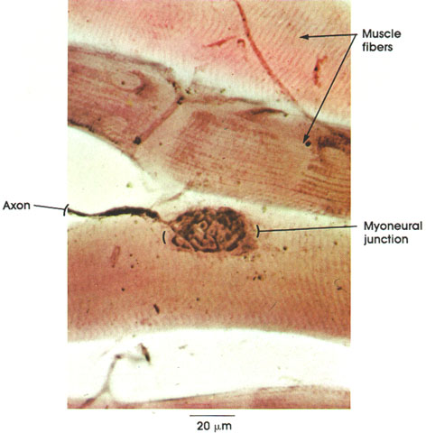
The method used in this preparation is a classical technique for staining nerve endings The muscle fibers seen here are not sectioned but are merely teased apart or spread by pressure. The advantages of this method are that it is simple and provides a broader view of the nerve terminals found on each muscle fiber in the preparation.
Muscle fibers: A small portion of three cross-striated muscle fibers is seen in this preparation.
Axon: A myelinated nerve fiber, upon reaching the muscle surface, loses its myelin sheath and branches extensively in a well-defined region called the motor end plate or myoneural junction.
Myoneural junction: The specialized region between the axon terminals and muscle fiber surface at which the nerve impulse is transmitted to the sarcolemma, resulting in muscular contraction.
Neural components
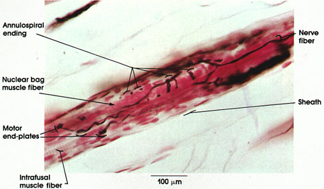
Muscle spindles are found within skeletal muscles. Each spindle is formed of 2 to 10 small muscle fibers, the intrafusal fibers, enclosed within a sheath of connective tissue that is pierced by nerve fibers.
Nerve fibers: Leaving and entering the muscle spindle, they may carry the sensory signals (output) or the motor signals (input).
Sheath: Connective tissue capsule that surrounds the intrafusal muscle fibers of the spindle.
Annulospiral endings: Also known as primary or nuclear bag endings. Large axon with many branches and terminal enlargements. Arborization of this type of ending occurs around the nuclear bag variety of intrafusal muscle fibers. These endings have a low threshold to stretch. They discharge when the intrafusal muscle fibers are stretched. The receptors are silent when the extrafusal (ordinary) muscle fibers contract and the intrafusal fibers are relaxed. Central processes of the annulospiral endings in the spinal cord participate in the monosynaptic (myotatic) reflex.
Nuclear bag muscle fibers: Larger variety of intrafusal muscle fibers. They have an enlarged equatorial region to accommodate numerous small nuclei. It is here that annulospiral endings arborize.
Motor end plates: The smaller nerve fibers within the spindle are axons of gamma neurons in the spinal cord. The axons terminate as typical motor end plates on intrafusal muscle fibers.
Intrafusal muscle fibers: Small striated muscle fibers rich in sarcoplasm and arranged parallel to the extrafusal skeletal muscle fibers. Two to 10 fibers enclosed in a connective tissue capsule form the muscle spindle.
Gastrocnemius muscle
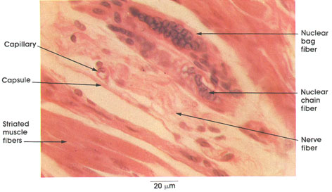
This specimen was obtained from a patient with a rhabdomyosarcoma, a very uncommon, highly malignant tumor of striated muscle. The tumor cells are not seen in this section.
Striated muscle fiber: Extrafusal (ordinary) skeletal muscle fiber oriented parallel to the intrafusal (encapsulated) fibers.
Capsule: Thin connective tissue sheath that surrounds the muscle spindle.
Nerve fiber: Myelinated axon. One of several nerve fibers related to the sensory and motor functions of the muscle spindle.
Nuclear chain fiber: Smaller variety of intrafusal muscle fibers (10 to 12 µm in diameter and 3 to 4 mm in length). Has a single row or chain of central nuclei.
Nuclear bag fiber: Larger variety of intrafusal muscle fibers. Enlarged equatorial region accommodates numerous small nuclei
Tendon of Achilles
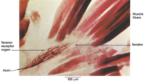
Axon: Myelinated axons break into primary, secondary, and tertiary branches. Unmyelinated branches from these axons wind around and in between tendon fascicles.
Tendon: Collagenous connective tissue bundle joined to several muscle fibers.
Muscle fibers: Skeletal (striated) muscle fibers.
Tension receptor organ (Golgi tendon organ): Discharges electrical impulses in response to tension on the tendon produced by either muscular contraction or muscle stretch. Provides information about the state of the muscle tension that determines in part the response of the central nervous system in the appropriate use of the muscle or muscles for precise motor function. Afferent impulses from Golgi tendon organs are conveyed to the central nervous system via lb nerve fibers, which exert presynaptic inhibition on the la nerve fibers originating in the muscle spindle.
Finger tip

Cynamologus monkey, glutaraldehyde-osmium,
toluidine blue, 854 x.
Meissner's* corpuscles are located in the dermal papillae of skin and are usually in contact with the basal cells of the epithelium. They are surrounded by a thin connective tissue sheath. Nerve terminals within the corpuscle are not seen with this method. Tactile corpuscles are common in hairless (glabrous) parts of the skin, but they are most numerous in the finger tips, palm of the hand, and sole of the foot.
Meissner's corpuscles are rapidly adapting mechanoreceptors that subserve discriminative touch sensations.
Dermal papillae are formed from the superficial dermis and are composed of collagenous connective tissue. The papillae contain, in addition to Meissner's corpuscles and their myelinated axons, so-called free nerve endings and capillary blood vessels, which, along with sweat glands, play an important role in temperature regulation in man.
Note the sweat gland duct within an epiderminal peg (ridge) and its coiled glandular portion, seemingly unattached, deep in the dermis.
.
(PACINIAN CORPUSCLE)
Pancreas
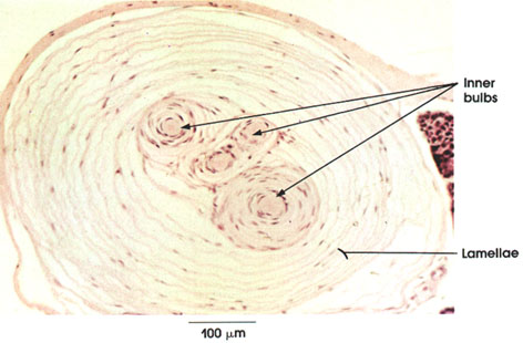
Pacinian corpuscles are mechanoreceptors found in the pancreas of cats but not man. In man and other animals, they are readily seen in sections of the dermis from the fingers and palm of the hand, the conjunctiva, near joints, in the mesenteries, branching blood vessels, penis, urethra, clitoris, parietal peritoneum, and loose connective tissue.
The Pacinian corpuscle is a pressure receptor and responds to high-frequency vibratory stimuli. Since the corpuscle is fluid-filled, it is essentially incompressible. The corpuscle transmits mechanical stimuli, through the connective tissue lamellae and fluid, to excite the nonmyelinated receptor axon in its core.
Pacinian corpuscles vary in size, but many are large enough to be easily dissected without magnifying lenses in the fingers of man.
Inner bulbs: Transverse section of branches of terminal unmyelinated nerve endings.
Lamellae: Concentric layers of collagenous connective tissue and flattened fibroblasts.
(CORPUSCLES OF VATER-PACINI)
Gross dissection
Shekleton's (ca. 1820) dissection of the digital branches of the median nerve demonstrating numerous, bulbous pacinian corpuscles is shown in this illustration. The illustration was published in 1848 in a book entitled Treatise on the Pathology, Diagnosis and Treatment of Neuroma, by R. W. Smith.
These end-organs are the only sensory receptors that are large enough to be identified and dissected in the anatomy laboratory. They should be looked for when dissecting the palm of the hand and fingers.
Tongue lamina propria
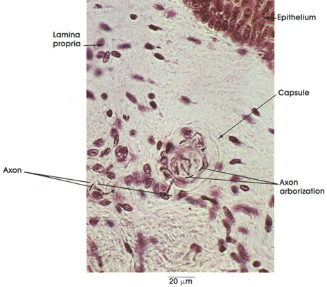
Axon: Myelinated sensory axon leading to the central nervous system.
Axon arborization: Encapsulated nonmyelinated terminal receptor branches of the sensory axon. They appear here as fine black lines within the capsule.
Sheath: Collagenous connective tissue spherical sheath or capsule enclosing the axon arborizations.
Connective tissue: Collagenous connective tissue of the lamina propria.
The end bulbs of Krause* are prevalent in mucous membranes and are believed to be cold temperature receptors
Penis ventral surface
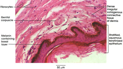
Beneath the thin stratified squamous keratinized epithelium, in the dense irregular connective tissue of the dermis, genital corpuscles may be found. These tactile-type sensory receptors are similar in appearance to Meissner's corpuscles except that they are not found in dermal papillae. They are found most frequently in the dermis on the ventral side of the penis.
Stimulation of these receptors leads to erection and ejaculation of semen.
Note that the cells of the deep layer of the skin contain melanin pigment granules
Protoplasmic astrocytes
Microglia
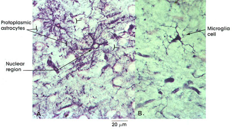
Protoplasmic astrocyte: It is stellate in shape, with many cytoplasmic processes. It is found chiefly in gray matter of brain and spinal cord and is important in metabolite transport.
Nuclear region: Nucleus of the protoplasmic astrocyte is unstained. It is large and rounded or ovoid.
Microglia cell: This is a small, dark cell. Processes fewer than those of the astrocyte, spiny and much more delicate. Microglia become phagocytes when cellular debris is present. Microglia are of mesodermal origin and migrate into the central nervous system and increase in number with nervous system damage
Fibrous astrocytes
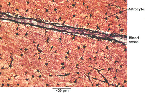
Astrocytes: Stellate cells. Processes fewer, straighter, and much longer than those of protoplasmic astrocytes .Note the relationship to blood vessels. Astrocyte processes with end feet are applied to the walls of blood vessels forming a continuous glial membrane surrounding blood vessels and capillaries. This important component of the so-called blood-brain barrier modifies the diffusion of substances from the blood to the extracellular fluid.
Oligodendroglia
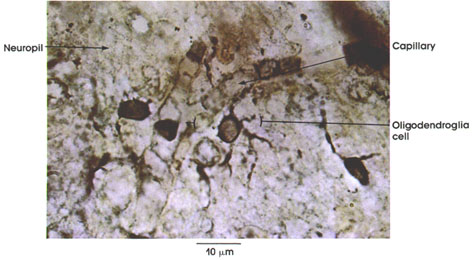
Oligodendroglia cell: Rounded nucleus, scanty cytoplasm, and a few delicate processes that extend for a short distance. Oligodendroglia are found in gray and white matter. They are usually seen in relation to neuronal cell bodies or between myelinated fibers. A cell of this type has also been described in juxtaposition to blood vessels. Oligodendroglia are believed to elaborate central nervous system myelin.
Neuropil: The region between neuronal and glial cells packed with neuronal and glial cell processes
Spinal cord
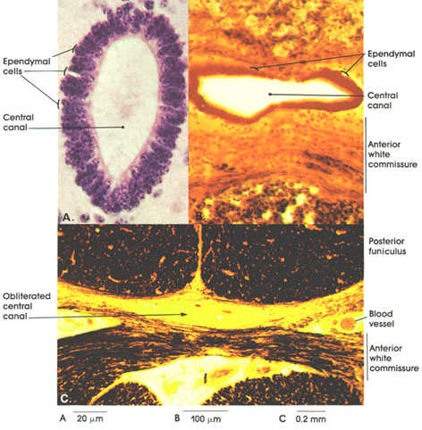
B. Bovine, Mü11er's fluid, Pal-Weigert and carmine stains, 162 x.
C. Cat, Mü11er's fluid, Pal-Weigert, 50 x.
The ependymal lining of the central canal of the spinal cord is shown in this plate. In A, note that the epenclymal cells are columnar in shape and are closely packed with their long axes perpendicular to the central canal. Their nuclei are elongated. Ependymal cells line the cavities of the spinal cord and brain (central canal and ventricles). Although the central canals seen (A and B) are patent, in adult humans and in some animals, the canal is usually obliterated (C). In B, note the anterior white commissure passing anterior (ventral) to the central canal. In C, in addition, note the posterior funiculus located posterior (dorsal) to the central canal
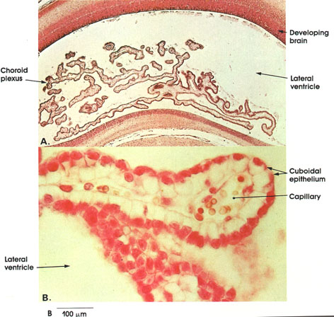
A: A low-magnification plate showing the branched projections of the choroid plexus within the cavity of the lateral ventricle in a developing cat brain.
B: A high-magnification plate showing the histology of the choroid plexus. Note the single layer of cuboidal epithelium with large spherical nuclei. Beneath the epithelium is a connective tissue core containing vascular channels. The choroid plexus is a major site for production of cerebrospinal fluid.